Understanding Common Salivary Gland Disorders and Their Warning Signs
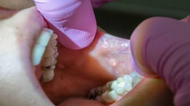
The human oral cavity is continuously lubricated and protected by a sophisticated network of salivary glands, consisting of three pairs of large major glands—the parotid, submandibular, and sublingual glands—alongside hundreds of microscopic minor glands scattered throughout the mucosal lining. Under healthy physiological conditions, these exocrine structures synthesize and secrete saliva, a complex fluid rich in water, electrolytes, antibacterial enzymes, and digestive proteins like amylase that facilitate mastication, initiate carbohydrate breakdown, preserve oral mucosal integrity, and maintain dental hygiene. Each major gland utilizes a specialized ductal system to channel saliva directly into the mouth. The parotid glands empty through Stensen's duct adjacent to the upper molars, the submandibular glands drain via Wharton's duct beneath the tongue, and the sublingual glands secrete through multiple minor ducts. When physical obstructions, ascending bacterial infections, autoimmune inflammatory attacks, or neoplastic transformations disrupt this secretory architecture, individuals face painful salivary gland disorders that impair basic oral functions.
Sialolithiasis: Pathophysiology of Salivary Duct Obstruction
One of the most frequent mechanical complications affecting the salivary glands is sialolithiasis, a condition characterized by the formation of calcified mineral stones—known as salivary stones or sialoliths—within the ductal system. These stones are composed primarily of calcium phosphate and calcium carbonate, along with organic debris, cellular fragments, and mucins. The submandibular gland is uniquely predisposed to stone formation, accounting for the vast majority of cases, largely due to anatomical and chemical factors: submandibular saliva possesses a more alkaline pH, a higher concentration of calcium and mucin, and flows upward against gravity through a long, tortuous duct known as Wharton's duct. When salivary stasis occurs, these mineral components precipitate around a microscopic nidus, gradually enlarging over time to partially or completely obstruct the ductal lumen, triggering painful swelling whenever salivary secretion is stimulated by the sight, smell, or taste of food.
Sialadenitis: Ascending Bacterial Infections and Inflammatory Responses
When salivary duct obstruction prevents normal salivary flow, or when systemic dehydration reduces overall fluid output, bacteria residing in the oral cavity can easily ascend backward through the ductal openings to colonize the glandular parenchyma, resulting in a painful infection known as sialadenitis. Acute bacterial sialadenitis typically affects the parotid gland in elderly, dehydrated, or debilitated patients, presenting as a rapid, tender swelling accompanied by localized erythema, skin warmth, and purulent discharge that can be expressed from the duct orifice upon manual massage. The predominant causative pathogen is Staphylococcus aureus, though other oral flora are frequently involved. In addition to acute bacterial infections, chronic recurrent sialadenitis can develop secondary to repeated ductal trauma or stricturing, leading to permanent acinar destruction, progressive fibrosis, and a persistent, dull ache within the affected gland.
Autoimmune Pathologies: Sjogren's Syndrome and Chronic Inflammation
Beyond mechanical stones and acute bacterial infections, the salivary glands are frequently targeted by chronic systemic autoimmune disorders, most notably Sjogren's syndrome. Sjogren's syndrome is a chronic inflammatory condition characterized by the progressive infiltration of autoreactive T-lymphocytes and B-lymphocytes into the exocrine glands, specifically targeting the lacrimal and salivary tissues. This immune-mediated attack destroys the acinar and ductal cells, severely diminishing salivary production and culminating in chronic xerostomia—a state of profound dry mouth that causes severe difficulties with swallowing, speaking, and maintaining dental health due to rampant tooth decay. The chronic lymphocytic infiltration also causes bilateral, painless enlargement of the major salivary glands, particularly the parotid glands, creating a distinct physical presentation that requires comprehensive serological testing and minor salivary gland biopsies to confirm the autoimmune diagnosis.
Salivary Gland Neoplasms: Benign Tumors and Malignant Transformations
The salivary glands can also give rise to a diverse spectrum of neoplastic growths, ranging from benign, slow-growing tumors to aggressive, locally destructive malignancies. Approximately eighty percent of all salivary gland tumors originate within the parotid glands, and the vast majority of parotid masses are benign, most commonly pleomorphic adenomas. A pleomorphic adenoma is a mixed tumor composed of epithelial and stromal elements that expands slowly over years without causing nerve damage. However, these benign lesions carry a well-documented risk of malignant transformation if left untreated for decades. Conversely, tumors arising in the smaller submandibular and sublingual glands carry a significantly higher statistical likelihood of being malignant. Common malignant salivary neoplasms include mucoepidermoid carcinoma, adenoid cystic carcinoma, and acinic cell carcinoma, all of which exhibit aggressive local invasion and a strong propensity to infiltrate nearby neural structures.
Critical Warning Signs and Clinical Symptom Presentation
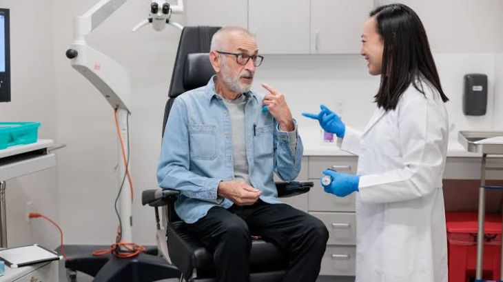
Recognizing the early warning signs of salivary gland pathology is essential for differentiating benign inflammatory conditions from dangerous malignancies that demand urgent intervention. Recurrent, painful swelling of the submandibular or parotid gland that intensifies dramatically immediately before or during meals, subsiding slowly afterward, is a classic warning sign of mechanical ductal obstruction caused by sialolithiasis. A critical clinical red flag in parotid gland tumors is the presence of facial nerve palsy or progressive weakness of the muscles of facial expression on the affected side. Because the facial nerve courses directly through the substance of the parotid gland, a tumor that infiltrates or compresses this nerve demands immediate oncological evaluation, as benign tumors virtually never cause nerve paralysis. Unexplained persistent pain, rapid tumor growth, fixation of the mass to deep underlying tissues or overlying skin, and palpable regional lymphadenopathy in the neck serve as vital warning signs indicating potential malignant transformation.
Diagnostic Investigations: Imaging and Histological Evaluation
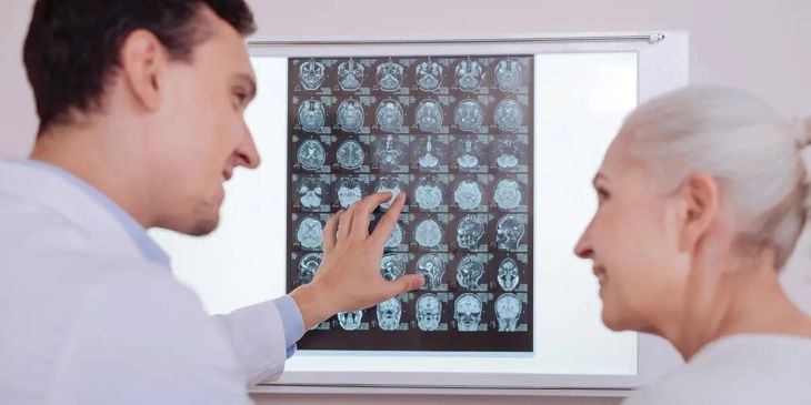
Evaluating complex salivary gland disorders requires a systematic diagnostic approach combining physical examination with advanced imaging and tissue sampling modalities. High-resolution ultrasonography serves as an excellent, non-invasive initial imaging tool to identify radiopaque salivary stones, evaluate ductal dilation, and differentiate between solid and cystic masses. When investigating deep-seated tumors or assessing local invasion into the skull base or bone, magnetic resonance imaging provides superior soft-tissue resolution, while computed tomography scans are ideal for detecting calcified stones and evaluating bony erosion. To establish a definitive histological diagnosis prior to surgical intervention, clinicians frequently perform a fine-needle aspiration biopsy, allowing pathologists to examine cellular architecture under a microscope and distinguish benign adenomas from aggressive carcinomas.
Therapeutic Interventions and Surgical Management
Managing salivary gland disorders requires targeted treatments tailored precisely to the underlying pathology, ranging from conservative medical management to complex surgical resections. Obstructive sialolithiasis is managed through conservative hydration, warm compresses, and sialogogues to stimulate salivary flow, though modern minimally invasive techniques like sialendoscopy now allow urologists and ENT specialists to thread tiny fiber-optic scopes directly into the ducts to laser or extract stones without open surgery. Acute sialadenitis requires targeted antibiotic therapy and gland massage, while chronic cases may necessitate surgical excision. For salivary gland neoplasms, surgical excision remains the gold standard of therapy. When operating on parotid tumors, surgeons perform meticulous superficial or total conservative parotidectomies designed to painstakingly dissect and preserve every delicate branch of the facial nerve, occasionally combining surgery with adjuvant radiation therapy for high-grade malignant carcinomas.
Conclusion
Salivary gland disorders encompass a wide spectrum of conditions—ranging from painful calcium stone obstructions and ascending bacterial infections to autoimmune Sjogren's destruction and aggressive carcinomas—that significantly impact oral health. By examining how ductal stasis, inflammation, and cellular mutations disrupt exocrine secretion, the critical importance of recognizing warning signs like meal-related swelling and facial nerve weakness becomes evident. Through precise ultrasound imaging, sialendoscopy, fine-needle aspiration biopsies, and nerve-sparing surgical interventions, healthcare providers can successfully restore salivary function, eliminate infection, and protect patients from malignant disease.