Normal Pressure Hydrocephalus How Does Affect Older Adults
The central nervous system is continuously bathed, cushioned, and nourished by a clear, ultrafiltered liquid known as cerebrospinal fluid, which circulates smoothly throughout the cranial cavity and spinal canal. This vital fluid is produced primarily by specialized vascular networks called the choroid plexuses residing inside the brain's interconnected ventricular system, consisting of the lateral ventricles, the third ventricle, and the fourth ventricle. Under normal physiological conditions, cerebrospinal fluid flows continuously from the lateral ventricles downward into the basal cisterns, spreads across the outer surface of the cerebral cortex within the subarachnoid space, and is ultimately reabsorbed into the venous bloodstream through microscopic arachnoid granulations. This balanced production and reabsorption cycle maintains stable intracranial pressure and shields delicate neural tissue from physical trauma. However, when the normal drainage pathways fail or fluid absorption becomes impaired later in life, cerebrospinal fluid accumulates abnormally inside the brain ventricles, giving rise to a distinct neurological condition known as normal pressure hydrocephalus.
Pathophysiology: Ventricular Enlargement and Intracranial Pressure Dynamics
To comprehend the complex nature of normal pressure hydrocephalus, one must analyze its unique and counterintuitive fluid dynamics. Unlike acute forms of hydrocephalus where fluid blockage causes soaring intracranial pressures that compress brain tissue rapidly, normal pressure hydrocephalus features a gradual, chronic accumulation of cerebrospinal fluid within the ventricles without a sustained elevation in opening pressure measured during lumbar punctures; the pressure typically remains within normal limits or fluctuates intermittently. As excess fluid collects inside the ventricular chambers, the ventricles expand outward, stretching and distorting adjacent white matter tracts—particularly the delicate periventricular nerve fibers responsible for controlling lower-limb coordination, bladder function, and cognitive processing. This chronic mechanical stretching disrupts neural connectivity and blood flow, resulting in progressive neurological decline that frequently mimics other neurodegenerative conditions common in older populations.
Primary Classifications: Idiopathic Versus Secondary Hydrocephalus
Clinicians divide normal pressure hydrocephalus into two distinct etiological classifications based on the root cause of the cerebrospinal fluid disturbance.
-
Idiopathic Normal Pressure Hydrocephalus: The vast majority of cases diagnosed in older adults are classified as idiopathic, meaning the precise underlying trigger remains unknown, though it is strongly associated with age-related changes in vascular compliance, impaired lymphatic clearance, and diminished arachnoid granulation reabsorption.
-
Secondary Normal Pressure Hydrocephalus: Conversely, secondary forms develop as a direct consequence of a known preceding neurological insult or injury that disrupts normal cerebrospinal fluid pathways. Common precipitating events include severe subarachnoid hemorrhage from a ruptured cerebral aneurysm, bacterial or viral meningitis, traumatic brain injury, or previous intracranial surgical procedures that leave behind inflammatory scarring and adhesions in the subarachnoid space.
The Classic Clinical Triad: Gait, Cognitive, and Urinary Symptoms
Normal pressure hydrocephalus presents with a classic constellation of clinical indicators historically known as Hakim's triad, comprising gait disturbance, cognitive impairment, and urinary incontinence.
-
Gait Disturbance: The earliest, most prominent, and often most debilitating feature is a characteristic gait disturbance, frequently described as a magnetic or shuffling walk where the patient's feet appear glued to the floor. Individuals exhibit a broad-based stance, reduced step height, impaired balance, and hesitation when initiating movement, greatly increasing their risk of frequent falls.
-
Cognitive Decline: Cognitive impairment manifests as a subcortical dementia characterized by executive dysfunction, slowed thinking speed, apathy, diminished attention span, and poor memory retrieval rather than primary aphasia.
-
Urinary Dysfunction: Urinary symptoms typically progress from initial urinary urgency and frequency to full-blown urge incontinence as the periventricular tracts controlling bladder sphincter tone are stretched and compressed by the expanding ventricles.
Diagnostic Investigations: Imaging and Lumbar Drainage Trials
Diagnosing normal pressure hydrocephalus in older adults requires a careful, multidisciplinary evaluation because its clinical presentation heavily overlaps with more common conditions like Alzheimer's disease, Parkinson's disease, or normal age-related mobility changes.
-
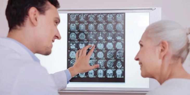
Neuroimaging Studies: Brain imaging via magnetic resonance imaging or computed tomography serves as the cornerstone of diagnosis, revealing disproportionate enlargement of the cerebral ventricles that cannot be explained solely by generalized brain shrinkage, accompanied by sharp callosal angles and tight high-concurrence sulci.
-
Diagnostic Tap and Lumbar Drainage: To confirm that the patient's symptoms will respond favorably to surgical intervention, clinicians perform diagnostic cerebrospinal fluid removal procedures, such as a high-volume lumbar puncture or the placement of an temporary lumbar drain. If the patient demonstrates a marked, measurable improvement in their gait speed and cognitive performance immediately following fluid drainage, the likelihood of a successful surgical outcome increases dramatically.
Surgical Interventions: Ventriculoperitoneal Shunting and Outcomes
When diagnostic evaluations confirm normal pressure hydrocephalus and demonstrate positive responses to fluid removal, the definitive, life-changing treatment involves surgical intervention to divert the excess cerebrospinal fluid away from the brain. The most common and effective surgical procedure is the insertion of a ventriculoperitoneal shunt. During this operation, a neurosurgeon places a fine, flexible catheter through a small burr hole in the skull directly into the lateral ventricle of the brain, tunneling the tubing subcutaneously down the neck and chest into the peritoneal cavity of the abdomen, where excess fluid is safely absorbed by the body. To prevent over-drainage caused by gravity changes when standing up, modern shunts utilize programmable valves that allow physicians to adjust the resistance precisely. When performed successfully in carefully selected candidates, shunting can dramatically reverse gait abnormalities, restore independence, and significantly improve overall quality of life.
Postoperative Management and Prognostic Considerations
Navigating the long-term care of older adults treated for normal pressure hydrocephalus requires vigilant neurological follow-up and monitoring for potential surgical complications. While gait disturbances typically show the most dramatic and rapid recovery following shunt placement, cognitive improvements can be more gradual and variable depending on the duration of symptoms prior to surgery and the presence of concurrent neurodegenerative pathologies like Alzheimer's plaque deposition. Potential complications of shunting include mechanical obstruction of the tubing, over-drainage leading to subdural hematomas, or localized shunt infections. By maintaining proactive clinical oversight, managing valve settings, and combining surgical treatment with physical therapy and rehabilitation exercises, healthcare providers can maximize functional recovery and help older adults regain their mobility and independence.
Conclusion
Normal pressure hydrocephalus is a treatable neurological disorder characterized by chronic cerebrospinal fluid accumulation and ventricular enlargement in older adults, resulting in the classic triad of gait instability, cognitive slowing, and urinary incontinence. By exploring how impaired fluid reabsorption stretches periventricular pathways, understanding the critical diagnostic value of large-volume lumbar taps, and recognizing the transformative benefits of ventriculoperitoneal shunt surgery, the clinical significance of this condition becomes clear. Through meticulous diagnostic evaluation and targeted surgical diversion, healthcare providers can successfully alleviate debilitating symptoms and restore vitality to affected individuals.