Understanding Brugada Syndrome and Its Effects on Heart Rhythm
The human heart relies on a synchronized electrical conduction system to coordinate muscular contractions and circulate blood across the circulatory system. In a healthy cardiovascular network, microscopic ion gates open and close in rapid sequence, generating the precise electrical impulses recorded on standard electrocardiograms. When microscopic channel defects disrupt this ionic choreography, life-threatening arrhythmias can arise even within structurally normal hearts. Brugada syndrome stands out as one of the most enigmatic channelopathies known to clinical cardiology, characterized by a vulnerability to ventricular tachyarrhythmias and sudden cardiac arrest. First described in medical literature during the early 1990s, this inherited arrhythmogenic condition predominantly strikes young, otherwise healthy adults, frequently manifesting during moments of deep rest, sleep, or febrile illness. Because the myocardium shows no overt physical scars, hypertrophic changes, or coronary artery blockages on standard ultrasound imaging, detecting the disorder demands specialized electrocardiographic awareness and targeted genetic screening. Grasping the underlying physiological mechanisms of Brugada syndrome, recognizing its signature electrophysiological markers, and uncovering common situational triggers empowers at-risk individuals and healthcare providers to take decisive preventive steps before sudden, catastrophic cardiac events unfold.
The Molecular Mechanism: Ion Channel Mutations and Disrupted Cardiac Action Potentials
At the cellular level, Brugada syndrome stems from dysfunctional ion channel operation within the membranes of ventricular cardiomyocytes, predominantly across the right ventricular outflow tract. The cardiac action potential relies on a balanced movement of sodium, calcium, and potassium ions across the cellular membrane. In roughly twenty to thirty percent of identified cases, the condition links directly to pathogenic variants within the SCN5A gene, which provides genetic instructions for creating the alpha-subunit of the cardiac voltage-gated sodium channel Nav1.5. These loss-of-function genetic mutations reduce inward sodium currents during phase zero and phase one of the cardiac action potential cycle. Consequently, the delicate balance between inward depolarizing currents and outward repolarizing potassium currents, notably the transient outward potassium current known as Ito, becomes skewed. Because the outer epicardial layer of the right ventricular outflow tract naturally harbors a denser concentration of Ito channels than the inner endocardium, the reduction in sodium influx creates a marked voltage gradient across the ventricular wall. This transmural dispersion of repolarization sets the biological stage for phase two re-entry, sparking disorganized, rapid electrical circuits that degrade normal contractions into polymorphic ventricular tachycardia or lethal ventricular fibrillation.
Clinical Presentation, Electrocardiographic Patterns, and Hidden Warning Signs
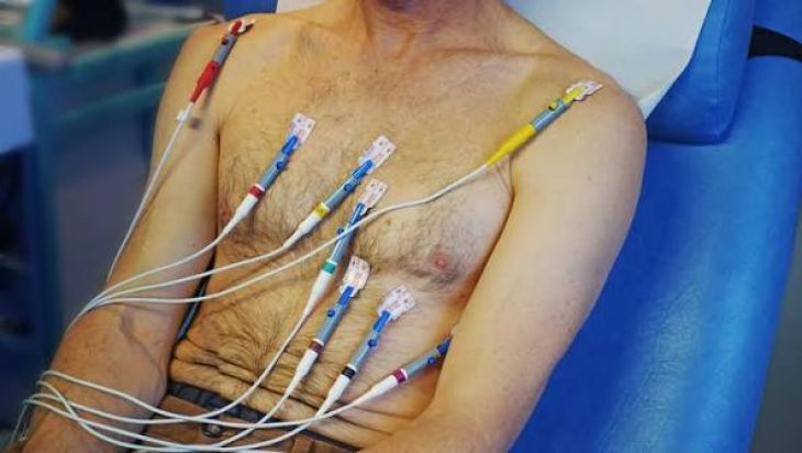
Brugada syndrome poses diagnostic challenges because many affected individuals remain completely asymptomatic until a sudden arrhythmic event occurs. When symptoms do emerge, they often appear as sudden unheralded syncope, where the individual faints abruptly without the warning lightheadedness or tunnel vision seen in ordinary vasovagal fainting. Other patients report sudden episodes of nocturnal agonal respiration, presenting as severe nighttime gasping, labored breathing, or seizure-like movements during sleep, which represent brief, self-terminating bouts of ventricular fibrillation. On a twelve-lead electrocardiogram, the hallmark diagnostic clue is the "Type 1" Brugada pattern observed in the right precordial leads (V1 through V3). This distinct tracing features a coved-type ST-segment elevation of two millimeters or more, followed immediately by a negative, inverted T-wave. A second manifestation, termed the "Type 2" or saddleback pattern, displays a high takeoff ST-elevation that slopes downward before rising into a biphasic or positive T-wave. Because these electrocardiographic abnormalities can fluctuate spontaneously over time—appearing clearly on one day and vanishing entirely on another—clinicians often must remain alert to subtle, intermittent changes during routine medical visits.
Environmental and Physiological Triggers That Unmask Arrhythmic Vulnerability
While Brugada syndrome is an inherited genetic condition, electrical destabilization often requires specific physiological triggers that unmask dormant channel dysfunction. High core body temperature is the most recognized clinical catalyst; fever alters the gating kinetics of mutant sodium channels, accelerating their inactivation and precipitating acute electrocardiographic changes and malignant arrhythmias. For this reason, pediatric and adult patients carrying the Brugada trait face heightened vulnerability during systemic viral or bacterial infections. Furthermore, autonomic tone exerts a distinct influence: arrhythmias occur most frequently at night or during deep rest, periods characterized by high vagal parasympathetic activity and reduced adrenergic stimulation. Certain dietary habits and chemical exposures also introduce risk, including heavy alcohol consumption, massive carbohydrate-heavy meals, and electrolyte imbalances such as hypokalemia or hyperkalemia. Most critically, an extensive list of pharmaceutical compounds can unmask the Brugada ECG pattern or provoke arrhythmias. These include Class IC antiarrhythmic agents like flecainide and ajmaline, local anesthetics, specific psychotropic medications like tricyclic antidepressants, and sodium-channel-blocking recreational substances, making strict pharmacological avoidance a central priority for diagnosed individuals.
Diagnostic Provocation, Risk Stratification, and Modern Treatment Paradigms
Confirming Brugada syndrome requires a multimodal diagnostic approach that combines baseline electrocardiography with targeted pharmacological provocation tests. When patients present with unexplained fainting or a family history of premature sudden death but show equivocal ECG tracings, electrophysiologists often administer intravenous sodium channel blockers, such as ajmaline, procainamide, or flecainide, in a monitored setting to uncover latent Type 1 patterns. High-resolution genetic testing complements this evaluation by identifying pathogenic variants in SCN5A and guiding family-wide cascade screening. Once confirmed, clinical risk stratification determines the appropriate therapeutic path based on prior syncope, documented ventricular arrhythmias, or spontaneous Type 1 presentations. For high-risk individuals who have survived sudden cardiac arrest or experienced arrhythmic syncope, the implantable cardioverter-defibrillator (ICD) represents the absolute standard of care, continually monitoring heart rhythm and delivering life-saving electrical shocks if ventricular fibrillation occurs. For patients with recurrent ICD shocks or those who are unsuitable candidates for defibrillator implantation, medical therapy using quinidine—which blocks the outward Ito current—helps stabilize electrical gradients. Additionally, catheter ablation targeting the arrhythmogenic substrate on the anterior epicardial surface of the right ventricular outflow tract offers an emerging, minimally invasive solution to suppress recurring electrical storms.
Conclusion: Proactive Management and Family Screening to Prevent Sudden Cardiac Death
Brugada syndrome highlights the delicate relationship between microscopic genetic architecture and whole-body cardiac stability, showing how a targeted ion channel defect can trigger sudden, life-threatening ventricular arrhythmias in an otherwise healthy heart. Because its clinical manifestations range from complete lifelong silence to sudden, nocturnal syncopal episodes, maintaining a high index of suspicion is essential when evaluating individuals with unexplained fainting or an inherited family history of unexplained young deaths. Contemporary cardiology provides robust strategies to manage this complex channelopathy, combining pharmacological drug challenge testing, targeted genetic analysis, and aggressive lifestyle modifications such as the rapid treatment of fevers and the strict avoidance of known channel-blocking medications. When paired with life-saving implantable cardioverter-defibrillators and modern epicardial catheter ablation techniques, at-risk patients can be effectively shielded from fatal rhythm disturbances. Through proactive medical guidance, comprehensive family cascade screening, and widespread public awareness, clinicians and affected families can demystify Brugada syndrome, prevent sudden cardiac arrest, and secure long-term, confident cardiovascular health.