Understanding Long QT Syndrome and Its Impact on Heart Rhythm
The human heart beats approximately one hundred thousand times every single day, quietly sustaining cellular life by delivering oxygenated blood across thousands of miles of vascular pathways. This mechanical miracle is driven by a microscopic electrical conduction system embedded directly within the myocardial tissue. Every coordinated contraction and subsequent relaxation phase depends on the precise movement of positively and negatively charged ions—principally sodium, potassium, and calcium—traveling back and forth across specialized cardiac cell membranes. These molecular gateways, known as ion channels, operate in split-second harmony to establish a stable and repetitive sinus rhythm.
When this finely tuned electrical timing experiences even slight disruptions, the heart's pumping efficiency can unravel rapidly. Among the most serious and clinically deceptive electrical channelopathies is Long QT Syndrome, frequently abbreviated as LQTS. Named after an extended diagnostic interval visible on an electrocardiogram tracing, this condition delays the critical period the heart ventricles require to electrically recharge between contractions. While many individuals harboring this condition remain unaware of its existence, a prolonged recharging phase creates an underlying vulnerability to chaotic and potentially lethal ventricular rhythms. Recognizing the underlying physiological mechanisms, clinical triggers, and therapeutic pathways associated with Long QT Syndrome is essential for preserving cardiovascular health and preventing catastrophic cardiac events.
What Is Long QT Syndrome and How Does the QT Interval Function?
To fully comprehend the mechanics of Long QT Syndrome, it is necessary to examine how electrical conduction is recorded using an electrocardiogram. A standard electrocardiogram translates the continuous cycle of cardiac polarization and repolarization into a sequence of distinct wave deflections labeled as the P, Q, R, S, and T waves. The initial P wave reflects atrial depolarization, signaling the upper chambers to squeeze blood downward into the ventricles. The prominent QRS complex represents the rapid depolarization of the muscular ventricular chambers, generating the forceful pressure needed to circulate blood throughout the body. Following this contraction, the T wave signifies ventricular repolarization, which is the electrical recovery period during which myocardial cells replenish their resting potentials before the next cycle begins.
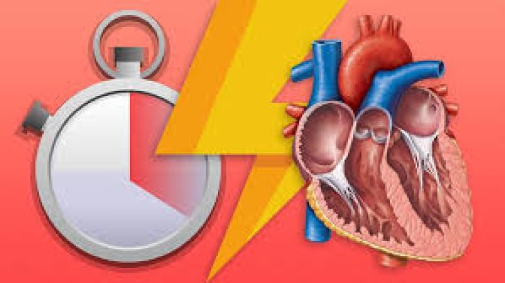
The QT interval itself measures the precise temporal duration spanning from the earliest onset of the Q wave to the complete baseline resolution of the T wave. This interval captures the cumulative time required for the lower cardiac chambers to activate and subsequently recover. Because human heart rates naturally speed up and slow down throughout the day in response to physical activity and emotional states, physicians standardize this measurement by calculating a rate-corrected QT interval, widely designated as the QTc. In healthy populations, normal QTc values generally remain below 440 milliseconds in adult men and 460 milliseconds in adult women. When this corrected value persistently stretches beyond 480 milliseconds, medical professionals begin evaluating for Long QT Syndrome.
During an abnormally elongated repolarization phase, the ventricular muscle remains in an electrically unstable, vulnerable refractory state. Under ordinary conditions, cardiac tissue cannot be easily re-excited until repolarization is finished. However, in Long QT Syndrome, specific localized clusters of heart cells finish recharging earlier than adjacent areas, creating substantial electrical heterogeneity across the heart wall. If a stray electrical impulse fires into this vulnerable refractory window, it can disrupt organized conduction and trigger rapid, self-sustaining circular electrical loops that render the heart unable to sustain a life-supporting pulse.
Congenital versus Acquired Long QT Syndrome: Origins and Root Causes
Long QT Syndrome is fundamentally categorized into two principal forms depending on its underlying etiology: congenital and acquired. The congenital variant arises from inherited genetic mutations that encode the structural proteins forming cardiac ion channels. Researchers have cataloged numerous distinct genetic subcategories, but the overwhelming majority of all congenital diagnoses belong to one of three primary classifications. Type 1 Long QT Syndrome involves mutations in the KCNQ1 gene, causing a deficiency in the slow delayed rectifier potassium current and creating vulnerability during rigorous physical exertion, especially swimming. Type 2 Long QT Syndrome is driven by KCNH2 gene mutations that disturb rapid potassium channels, making individuals exceptionally reactive to sudden auditory frights or acute emotional shocks. Type 3 Long QT Syndrome stems from SCN5A mutations that impair the prompt closing of sodium channels, causing an abnormal inward trickle of positive ions that frequently sparks rhythm disruptions during deep resting states or nighttime sleep.
Conversely, acquired Long QT Syndrome develops much later in life and occurs independently of inherited DNA defects, though certain individuals may carry a mild genetic predisposition that only emerges when exposed to external pharmacological or metabolic stressors. The most common trigger for the acquired variant is the administration of prescription medications that inadvertently block the human ether-a-go-go-related gene potassium channel. Hundreds of widely used drugs have been identified as potential culprits, including specific class IA and class III antiarrhythmic agents, macrolide and fluoroquinolone antibiotics, certain antipsychotics, selective serotonin reuptake inhibitors, and common anti-nausea preparations. When these medications accumulate in the bloodstream, they systematically prolong ventricular repolarization.
In addition to pharmaceuticals, severe disruptions in serum electrolyte concentrations frequently precipitate acquired Long QT Syndrome. Ion channels depend on precise biochemical concentrations in the extracellular fluid to preserve membrane gradients. Marked drops in circulating potassium or magnesium levels, often brought on by aggressive diuretic therapy, prolonged bouts of vomiting, severe gastrointestinal illness, or advanced eating disorders, drastically hinder potassium outflow from myocardial cells. Severe clinical hypothyroidism, heart failure, and extreme starvation can likewise weaken cardiac repolarization kinetics, producing dangerous shifts in the QT interval.
The Pathophysiological Impact: Arrhythmogenesis and Torsades de Pointes
The core physiological threat posed by Long QT Syndrome is its capacity to trigger catastrophic ventricular tachyarrhythmias. When ventricular repolarization is prolonged, myocardial cells become susceptible to early afterdepolarizations, which are abnormal spontaneous depolarizing spikes that arise before the cell membrane has fully repolarized. If one of these premature depolarizations coincides precisely with the crest or descending slope of the preceding T wave, an electrophysiological event known as the R-on-T phenomenon occurs. This rogue excitation acts like a sudden spark within a chamber of volatile gas, fracturing normal cardiac rhythm into continuous electrical turbulence.
The specific arrhythmia most characteristically tied to Long QT Syndrome is known clinically as Torsades de Pointes, a descriptive term derived from the French phrase for twisting of the points. On an electrocardiographic monitor, this polymorphic ventricular tachycardia appears as a series of undulating waveforms that continuously twist their amplitude and electrical axis around the central baseline. During an episode of Torsades de Pointes, the ventricles beat erratically at speeds ranging between two hundred and two hundred and fifty cycles per minute. At this extreme velocity, the lower chambers lose their ability to relax, fill with venous blood, or generate effective mechanical contractions, leading to a sudden, dramatic plunge in arterial blood pressure.
When an episode of Torsades de Pointes lasts for only a few seconds before spontaneously terminating, the affected individual typically experiences sudden dizziness or transient loss of consciousness, often regaining alertness quickly once a regular rhythm resumes. However, if the chaotic electrical circus movements fail to stop on their own, the arrhythmia frequently degenerates directly into ventricular fibrillation. In ventricular fibrillation, coordinated myocardial contraction ceases entirely, the heart merely quivers, and systemic circulation stops completely. Without immediate emergency intervention utilizing cardiopulmonary resuscitation and an external defibrillator, this progression culminates in irreversible cerebral damage and sudden cardiac death within minutes.
Clinical Presentation, Diagnostic Evaluation, and Treatment Paradigms
Recognizing Long QT Syndrome can be remarkably challenging because a substantial portion of affected individuals display no day-to-day symptoms and lead seemingly healthy lives. When clinical manifestations do emerge, the hallmark indicator is unexplained syncope, which presents as an abrupt loss of consciousness without the typical prodromal sensations of nausea, warmth, or visual tunnel vision that usually precede benign fainting. These syncopal episodes frequently occur during athletic competition, intense mental distress, or sudden auditory stimulation. Additionally, the cerebral hypoperfusion that accompanies temporary cardiac arrest is frequently accompanied by involuntary muscle twitching and incontinence, causing many individuals with undiagnosed Long QT Syndrome to be incorrectly treated for neurological seizure disorders.
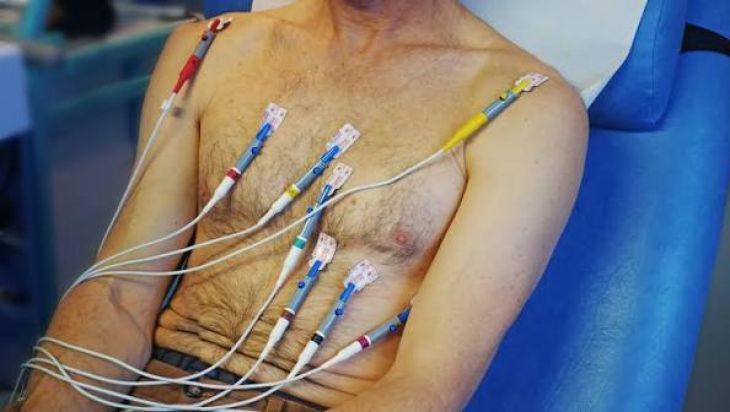
Accurate diagnosis requires an integrative clinical evaluation combining baseline electrocardiography with specialized secondary assessments. A standard resting twelve-lead electrocardiogram serves as the initial screening tool, although normal baseline readings can occasionally occur due to fluctuating autonomic tone. To unmask latent repolarization abnormalities, cardiologists frequently utilize treadmill exercise stress testing, continuous ambulatory Holter monitoring, and comprehensive genetic sequencing panels. Furthermore, clinicians often apply the Schwartz scoring criteria, an objective algorithmic framework that scores electrocardiographic findings, personal fainting histories, and family histories of sudden premature death to determine the diagnostic likelihood of Long QT Syndrome.
Once a diagnosis is firmly established, treatment strategies prioritize dampening sympathetic cardiac triggers and preventing lethal arrhythmias. Pharmacotherapy forms the foundation of clinical management, with non-selective beta-blockers such as nadolol and propranolol serving as first-line agents to shield the heart from adrenaline spikes during stress or exertion. For patients carrying sodium channel abnormalities, specialized channel blockers like mexiletine are frequently added to shorten the repolarization phase. When medical therapy proves inadequate, or in high-risk patients who have survived cardiac arrest, the surgical implantation of an internal cardioverter-defibrillator provides an essential safety net by delivering an immediate electrical shock if life-threatening arrhythmias emerge. Complementing these medical measures, patients must strictly avoid known QT-prolonging pharmaceuticals, maintain balanced hydration and electrolyte levels, and adopt lifestyle modifications appropriate for their specific genetic variant.
Conclusion: Living Safely and Confidently with an Electrical Heart Disorder
Long QT Syndrome stands as a clear reminder of how microscopic ion-channel irregularities can profoundly impact the mechanical rhythm of the human heart. Although the potential consequences of prolonged cardiac repolarization are severe, modern electrophysiology has transformed what was once a mysterious and fatal disorder into a manageable condition.
Early clinical identification remains the single most important factor in altering patient outcomes. Through widespread electrocardiographic screening, comprehensive family pedigree analysis, targeted pharmacotherapy, and implantable device integration, the overwhelming majority of people living with Long QT Syndrome can safely mitigate their risks. By pairing regular medical oversight with a clear understanding of personal triggers, individuals diagnosed with this condition can successfully protect their cardiovascular health and lead full, active, and confident lives.