What Is Spontaneous Coronary Artery Dissection and How Can It Affect the Heart?
Introduction to Non-Atherosclerotic Coronary Disruption
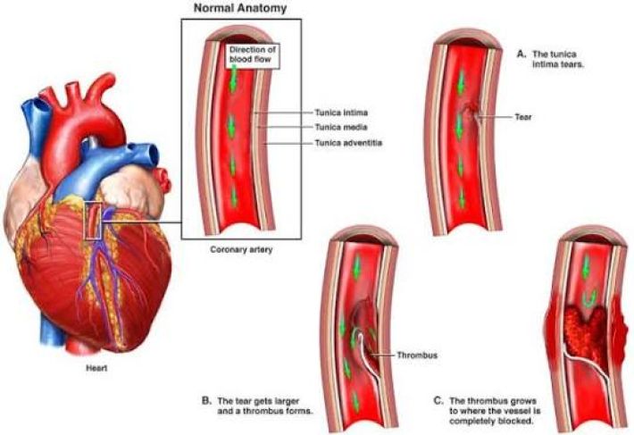
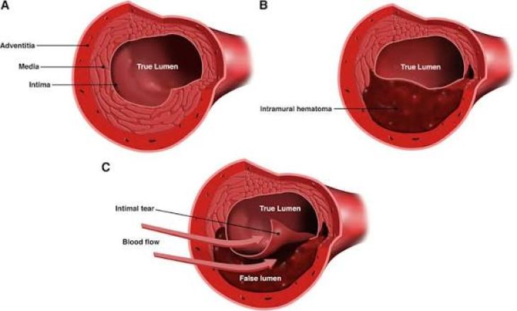
Continuous oxygenation of the cardiac muscle depends entirely upon the structural integrity of the coronary arterial tree. While the vast majority of acute coronary events stem from classic atherosclerotic plaque instability, Spontaneous Coronary Artery Dissection (SCAD) represents a distinct, non-atherosclerotic cause of acute myocardial ischemia and sudden cardiac events. This condition develops when a spontaneous structural breach or physical separation occurs within the tunica layers of a coronary vessel. Rather than originating from chronic endothelial lipid accumulation and calcification, this pathology involves an acute splitting of the vascular wall. This creates a false lumen that compresses or completely shuts off blood transit through the true arterial channel. SCAD predominantly affects young and middle-aged individuals—particularly females lacking traditional cardiovascular risk factors—presenting unique diagnostic and therapeutic considerations.
Histological Architecture and Mechanisms of Wall Separation
Understanding the physical dynamics of SCAD requires examining the trilaminar structure of coronary arteries, which consist of the innermost intima, the muscular media, and the outer adventitia. Vessel wall disruption proceeds via two primary mechanical pathways. The first involves a primary intimal tear, where a breach in the endothelial lining allows blood under high systemic pressure to penetrate the medial layer, creating an expanding secondary channel. The second pathway involves primary vasa vasorum hemorrhage, where microvascular blood vessels supplying the outer vessel wall rupture directly inside the medial layer without a preceding intimal breach. In both scenarios, localized fluid pooling forms an intramural hematoma. As this hematoma expands under pressure, it compresses the true arterial lumen, causing downstream tissue ischemia or complete vascular occlusion.
Predisposing Arteriopathies and Acute Stress Triggers
Arterial wall disruption rarely develops in completely normal vascular tissue; rather, it typically occurs where underlying structural arteriopathies intersect with acute physical or emotional stress. The most frequent predisposing vascular condition is fibromuscular dysplasia, a non-atherosclerotic, non-inflammatory disease that structurally weakens arterial walls. Systemic hormonal fluctuations also contribute significantly; elevated levels of progesterone and estrogen during pregnancy or the early postpartum state alter collagen and mucopolysaccharide composition, reducing overall vascular elasticity. Furthermore, inherited connective tissue disorders—including Marfan syndrome, vascular Ehlers-Danlos syndrome, and Loeys-Dietz syndrome—substantially increase vessel wall vulnerability. These structural predispositions are frequently unmasked by acute physical or emotional triggers, such as intense isometric exertion or severe emotional distress, which provoke sudden spikes in systemic blood pressure and arterial shear stress.
Ischemic Impacts on Myocardial Performance and Clinical Signs
The immediate physiological consequence of internal vessel layer separation is acute myocardial ischemia or frank tissue infarction resulting from restricted downstream perfusion. The clinical presentation mirrors that of traditional atherosclerotic acute coronary syndromes. Most affected individuals present with acute substernal chest pressure or crushing pain radiating to the arm, neck, or jaw, accompanied by dyspnea, diaphoresis, nausea, and lightheadedness. Electrophysiologically, ischemic cardiac tissue can precipitate severe ventricular dysrhythmias, including sustained ventricular tachycardia or ventricular fibrillation, leading to sudden circulatory collapse. The extent of functional myocardial impairment depends on the specific vessel involved—most commonly the left anterior descending artery—and how rapidly true luminal perfusion is compromised or restored.
Angiographic Classification and Intravascular Imaging
Accurate diagnostic identification during acute presentation is critical, as standard invasive interventions designed for atherosclerotic heart attacks can exacerbate vessel wall separation. While blood tests confirm elevated cardiac troponins and electrocardiograms reveal ischemic ST-segment shifts, definitive diagnosis requires invasive coronary angiography. Angiographically, the disease manifests in distinct patterns: Type 1 demonstrates classical contrast dye split with a visible dual lumen; Type 2 displays long, smooth arterial narrowing caused by contained intramural hematoma; and Type 3 mimics focal atherosclerotic stenosis. When angiographic findings are equivocal, advanced intravascular imaging modalities—such as Intravascular Ultrasound (IVUS) or Optical Coherence Tomography (OCT)—are utilized to cross-sectionally visualize the arterial wall, confirming the presence of intramural hematoma and false lumen boundaries.
Management Protocols and Long-Term Clinical Strategies
Therapeutic strategies for SCAD differ fundamentally from standard atherosclerotic coronary care. In hemodynamically stable patients without ongoing severe ischemia, conservative medical management serves as the preferred initial approach. Clinical observation demonstrates that the vast majority of dissected vessel segments undergo spontaneous healing over time when left unmanipulated. Conservative drug therapy typically involves antiplatelet agents to prevent thrombus formation within the true lumen, beta-blockers to decrease heart rate and reduce arterial shear stress, and statins only when concurrent hyperlipidemia is present. Invasive revascularization via Percutaneous Coronary Intervention (PCI) carries a high risk of iatrogenic complications, as balloon inflation or stent placement can extend the intramural hematoma proximally or distally; PCI is strictly reserved for patients with persistent hemodynamic instability, severe left main involvement, or refractory ischemia. Emergency Coronary Artery Bypass Grafting (CABG) remains a rescue option for extensive or complicated presentations.
Conclusion
Spontaneous Coronary Artery Dissection is a significant cause of non-atherosclerotic myocardial infarction driven by intramural hematoma formation and vessel wall separation. By understanding how underlying vascular conditions like fibromuscular dysplasia interact with acute physiological stress to cause luminal compression, the biological mechanisms governing acute cardiac ischemia become clear. Combining accurate angiographic classification with intravascular imaging enables clinicians to distinguish SCAD from traditional plaque rupture. Prioritizing conservative medical stabilization over routine stenting allows the injured arterial wall to heal naturally, minimizing procedural risks and preserving long-term myocardial performance.