What Is Lichen Planus and How Can It Affect the Skin and Mouth?
The human integumentary system and oral mucosal membranes rely on tightly regulated epithelial renewal and intact dermo-epidermal junctions to maintain mechanical protection, fluid balance, and immunological barrier defense. When an aberrant cell-mediated immune response targets the basal layer of stratified squamous epithelium, localized interface inflammation disrupts tissue architecture across both cutaneous and mucosal surfaces. Lichen Planus (LP) is a chronic, idiopathic, immune-mediated mucocutaneous inflammatory disorder that affects the skin, oral and genital mucous membranes, scalp, and nails. Most commonly presenting in middle-aged adults between thirty and sixty years of age, the condition exhibits distinct clinical morphologies depending on the anatomical site involved—ranging from intensely pruritic cutaneous papules to painful, erosive oral ulcerations. Understanding how lichen planus damages epidermal and mucosal tissues requires analyzing its cytotoxic T-lymphocyte immunopathology, basal keratinocyte apoptosis cascades, characteristic cutaneous and oral clinical patterns, histopathological hallmarks, and targeted anti-inflammatory therapies.
Immunopathological Mechanisms and Basal Keratinocyte Apoptosis
Although the precise initiating antigen remains unidentified, extensive immunohistological and molecular evidence demonstrates that lichen planus is driven by an antigen-specific, T-cell-mediated autoimmune assault against basal keratinocytes. Environmental or pharmacological triggers—including viral infections such as Hepatitis C virus, dental restorative materials like mercury amalgam, and systemic medications including angiotensin-converting enzyme inhibitors, beta-blockers, and nonsteroidal anti-inflammatory drugs—are thought to alter self-peptides expressed on basal epidermal cells. Antigen-presenting Langerhans cells process these modified surface antigens and present them via major histocompatibility complex (MHC) class I and class II molecules to naive T-lymphocytes within regional lymph nodes. Activated, clonally expanded cytotoxic CD8-positive T-cells and helper CD4-positive T-cells subsequently migrate back to the dermal-epidermal or lamina propria-epithelial junction, guided by upregulated vascular adhesion molecules and chemokines such as CXCL9 and CXCL10. Once positioned along the basal layer, cytotoxic CD8-positive T-lymphocytes trigger programmed cell death in basal keratinocytes through two synergistic pathways: exocytosis of cytolytic granules containing perforin and granzyme B, and engagement of Fas ligand (CD95L) on T-cells with Fas death receptors (CD95) on keratinocytes, activating intracellular caspase cascades.
Cutaneous Manifestations and the Koebner Phenomenon
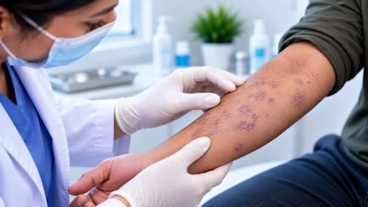
On keratinized skin, classic cutaneous lichen planus presents with a distinctive constellation of clinical features traditionally summarized by dermatologists using the mnemonic of the "six Ps": planar (flat-topped), purple (violaceous), polygonal, pruritic papules and plaques. These lesions typically measure two to ten millimeters in diameter and emerge symmetrically on the flexor surfaces of the wrists, forearms, ankles, dorsal hands, and lower back. Close inspection of the shiny, flat-topped papule surface often reveals fine, lacy white lines known as Wickham striae, which correspond histologically to focal zones of hypergranulosis. Pruritus is frequently severe and disproportionate to the visible rash, leading patients to rub rather than scratch their skin, which paradoxically avoids excoriation marks while inducing hyperpigmentation. A defining clinical hallmark of active cutaneous lichen planus is the Koebner isomorphic response, wherein new, linear arrays of characteristic violaceous papules erupt directly along sites of minor mechanical skin trauma, such as scratches, surgical incisions, or friction burns. Beyond classic papular eruptions, cutaneous variants include hypertrophic lichen planus (thick, hyperkeratotic plaques on the anterior shins), follicular lichen planopilaris (causing cicatricial scarring alopecia on the scalp), and nail lichen planus (causing longitudinal ridging, pterygium formation, and permanent nail plate loss).
Oral Mucous Membrane Involvement and Clinical Variants
Mucosal involvement occurs in roughly half of all patients with cutaneous lichen planus and frequently manifests as an isolated chronic condition without skin lesions. Oral Lichen Planus (OLP) exhibits a pronounced female predominance and tends to follow a more persistent, relapsing course than self-limiting cutaneous eruptions. Clinically, oral lichen planus is categorized into six distinct morphological subtypes that often coexist within the same oral cavity: reticular, papular, plaque-like, atrophic, erosive, and bullous. The reticular form is the most prevalent and typically asymptomatic, characterized by painless, bilateral, symmetrical networks of raised white, lace-like Wickham striae distributed across the posterior buccal mucosa, tongue, and gingiva. Conversely, atrophic and erosive oral lichen planus represent severe, symptomatic presentations caused by extensive basal cell destruction and complete epithelial detachment. Erosive lesions appear as irregular, fiery-red erythematous patches covered by yellowish fibrinous pseudomembranes, commonly affecting the buccal mucosa, lateral tongue borders, and gingiva—where diffuse erythema and sloughing produce desquamative gingivitis. Patients with erosive oral lesions suffer severe burning pain exacerbated by acidic, spicy, or coarse foods, impairing nutrition and oral hygiene. Furthermore, chronic erosive oral lichen planus carries a small but clinically significant risk of malignant transformation into oral squamous cell carcinoma, necessitating lifelong surveillance.
Histopathological Hallmarks and Diagnostic Confirmation
While classic cutaneous papules or symmetrical buccal Wickham striae can often be recognized clinically, atypical, hypertrophic, or erosive presentations require tissue biopsy for definitive diagnosis and to exclude mimicking conditions such as lupus erythematosus, pemphigus vulgaris, mucous membrane pemphigoid, and oral epithelial dysplasia. Histopathological examination of a lesional punch biopsy reveals a classic triad of interface dermatitis features. First, the epidermis demonstrates compact orthokeratosis, wedge-shaped hypergranulosis underlying Wickham striae, and irregular acanthosis that reshapes the rete ridges into a characteristic pointed "sawtooth" configuration. Second, the basal cell layer exhibits liquefactive (hydropic) degeneration accompanied by scattered, rounded eosinophilic apoptotic keratinocytes termed Civatte bodies (or colloid bodies) along the dermo-epidermal junction. Third, the superficial papillary dermis contains a dense, sharply demarcated, band-like lymphocytic infiltrate hugging the dermal-epidermal border without extending deep into the reticular dermis. Direct immunofluorescence microscopy provides supportive differentiation by revealing shaggy fibrinogen deposition along the basement membrane zone and immunoglobulin M staining within Civatte bodies, distinguishing lichen planus from autoimmune vesiculobullous disorders.
Therapeutic Strategies and Long-Term Disease Management
Management of lichen planus focuses on suppressing T-cell-mediated interface inflammation, alleviating pruritus and mucosal pain, and monitoring chronic mucosal lesions for dysplastic changes. While uncomplicated cutaneous lichen planus often resolves spontaneously within twelve to twenty-four months—leaving behind stubborn post-inflammatory hyperpigmentation—active symptomatic lesions respond wellto super-potent topical corticosteroids (such as clobetasol propionate) applied under occlusion, supplemented by oral antihistamines or narrowband ultraviolet B (NB-UVB) phototherapy for widespread disease. Severe, refractory cutaneous eruptions may require short tapering courses of systemic corticosteroids or oral retinoids like acitretin. For symptomatic erosive oral lichen planus, high-potency topical corticosteroid gels or rinses (such as fluocinonide or dexamethasone elixirs) serve as first-line therapy, while topical calcineurin inhibitors (tacrolimus or pimecrolimus) provide effective steroid-sparing maintenance for chronic mucosal erosions.
Conclusion
Lichen Planus is a chronic, T-cell-mediated mucocutaneous autoimmune disorder driven by cytotoxic CD8-positive lymphocyte destruction of basal keratinocytes along epithelial interfaces. Understanding how fas-mediated apoptosis and cytolytic granule release disrupt epidermal and mucosal integrity clarifies the clinical spectrum spanning pruritic violaceous cutaneous papules with Wickham striae to painful erosive oral ulcerations. Utilizing targeted tissue biopsies to identify sawtooth rete ridges, Civatte bodies, and band-like lymphocytic infiltrates enables clinicians to confirm the diagnosis and rule out premalignant dysplasia. Through potent topical corticosteroids, calcineurin inhibitors, phototherapy, and regular oral mucosal surveillance, multidisciplinary medical and dental teams can control inflammation, relieve debilitating symptoms, and preserve long-term tissue integrity.