What Is Aortic Dissection and Why Is It a Medical Emergency?
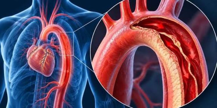
As the primary conduit emerging from the left ventricular outflow tract, the human aorta distributes oxygenated arterial blood across every systemic vascular bed while absorbing immense kinetic energy with each cardiac cycle. To endure repetitive pulsatile pressure waves without structural failure, the aortic wall relies on three distinct concentric histological coats: the inner tunica intima, the robust middle tunica media, and the protective outer tunica adventitia. Lining the vessel lumen, the tunica intima provides a smooth, non-thrombogenic endothelial surface anchored to a delicate basement membrane. Beneath it, the tunica media provides viscoelastic recoil and tensile durability via tightly organized lamellar units composed of smooth muscle cells, collagen fibrils, and fenestrated elastin sheets. Encasing the exterior, the tunica adventitia supplies mechanical tethering and houses the vasa vasorum, a network of penetrating microvessels that perfuse the outer medial layers. Aortic Dissection is a hyper-acute, life-threatening vascular emergency triggered when a sudden breach in the tunica intima permits high-pressure arterial blood to surge directly into the underlying tunica media. This pressurized column of blood cleaves the weakened medial lamellae apart longitudinally, carving out a secondary blood-filled channel known as a false lumen parallel to the native true lumen. Comprehending why aortic dissection demands immediate clinical intervention requires examining the biomechanical forces driving medial degeneration, rapid longitudinal propagation, vital organ malperfusion, and catastrophic free rupture.
Biomechanical Triggers and Structural Wall Degeneration
An intimal entry tear occurs when localized hemodynamic shear forces exceed the cohesive tensile strength of the aortic wall. Chronic systemic arterial hypertension serves as the dominant modifiable driver, underlying roughly three-quarters of all acute presentations. Persistent elevated blood pressure—especially steep acceleration of pressure during early ventricular ejection—exerts relentless cyclical stress against the inner lining while simultaneously compressing the vasa vasorum, inducing chronic ischemic atrophy of medial smooth muscle cells. Inherited connective tissue disorders accelerate a pathological process termed cystic medial degeneration, marked by fragmentation of elastic fibers, smooth muscle depletion, and pooling of mucopolysaccharide ground substance. Genetic syndromes such as Marfan syndrome (stemming from fibrillin-1 mutations), Loeys-Dietz syndrome, and vascular Ehlers-Danlos syndrome predispose patients to catastrophic wall failure early in adulthood. Similarly, a congenital bicuspid aortic valve disrupts laminar systolic ejection jets, directing abnormal helical shear stress against the ascending aortic wall and promoting progressive aneurysmal thinning. Acute triggers also include sympathomimetic surges from cocaine or amphetamine intoxication, severe deceleration chest trauma, late-pregnancy hemodynamic volume expansion, and mechanical catheter injury during endovascular procedures.
Pathophysiology of False Lumen Expansion and Organ Malperfusion
Once blood penetrates an intimal breach—typically located in the ascending aorta just above the sinotubular junction or immediately distal to the left subclavian artery—systolic pressure drives longitudinal separation along cleavage planes within the media. With every ventricular contraction, the pressurized false lumen propagates distally toward the iliac bifurcation and proximally toward the aortic root. This rapid expansion generates three lethal clinical mechanisms that explain why untreated proximal dissection carries a mortality rate climbing roughly one to two percent per hour after symptom onset. First, external rupture through the thin remaining tunica adventitia produces instantaneous, fatal hemorrhage into the pleural cavity, mediastinum, or pericardial sac; blood accumulating rapidly inside the closed pericardial space compresses the cardiac chambers, triggering acute cardiac tamponade and obstructive shock. Second, retrograde extension into the aortic root detaches the aortic valve commissures, precipitating acute, wide-open aortic regurgitation and fulminant cardiogenic pulmonary edema. Third, high pressure inside the false lumen compresses the true lumen or shears across the origins of vital arterial branches, inducing acute end-organ malperfusion—resulting in myocardial infarction when coronary ostia are occluded, hemispheric stroke when carotid perfusion fails, mesenteric bowel necrosis, or oliguric renal failure.
Anatomical Classification Systems and Clinical Triage
Urgent therapeutic triage in acute aortic dissection hinges on precise anatomical mapping, organized primarily through the Stanford and DeBakey classification frameworks. The Stanford system categorizes dissections into two practical groups dictated strictly by whether the ascending aorta is affected, irrespective of where the primary intimal tear originated. Stanford Type A dissections involve the ascending aorta—either limited to the proximal segment or extending across the aortic arch into the descending aorta—and mandate emergent open-heart surgical reconstruction due to imminent risks of pericardial tamponade, coronary occlusion, and free rupture. Stanford Type B dissections are confined strictly to the descending aorta distal to the left subclavian artery takeoff, sparing the ascending segment entirely. Providing further anatomical granularity, the DeBakey system delineates three patterns: DeBakey Type I begins in the ascending aorta and extends continuously past the arch into the descending aorta; DeBakey Type II remains isolated strictly within the ascending aorta; and DeBakey Type III originates in the descending thoracic aorta, subclassified into Type IIIa (ending above the diaphragm) and Type IIIb (propagating downward into the abdominal aorta).
Clinical Manifestations and Rapid Diagnostic Imaging
Individuals suffering acute aortic dissection characteristically experience the abrupt, instantaneous onset of severe, ripping or tearing pain centered in the anterior chest for Type A lesions or the interscapular upper back for Type B lesions. In contrast to angina pectoris, which typically escalates gradually, dissection pain achieves maximum intensity immediately at onset and frequently migrates downward as the medial cleavage plane advances along the trunk. Physical assessment often uncovers marked hemodynamic instability, severe hypertension or paradoxical hypotension signaling cardiac tamponade, systolic blood pressure discrepancies exceeding twenty millimeters of mercury between upper extremities, diminished or absent peripheral pulses, and an acute diastolic decrescendo murmur reflecting aortic valve incompetence. Swift, accurate diagnostic imaging is essential prior to invasive intervention. Contrast-enhanced Computed Tomography Angiography (CTA) spanning the thorax, abdomen, and pelvis represents the gold-standard diagnostic study in hemodynamically stable patients, delineating the undulating intimal flap separating true and false channels, localizing entrytears, and evaluating visceral branch perfusion. For critically unstable patients requiring immediate bedside evaluation, Transesophageal Echocardiography (TEE) rapidly visualizes the proximal ascending aorta, aortic valve integrity, and pericardial fluid accumulation.
Emergency Pharmacological Stabilization and Operative Repair
Initial therapy for acute aortic dissection centers on immediate intravenous anti-impulse medication aimed at blunting left ventricular ejection force and lowering mean arterial pressure to arrest further false lumen propagation. Short-acting intravenous beta-adrenergic antagonists—such as esmolol or labetalol—must be infused first to reduce heart rate below sixty beats per minute and diminish systolic pressure velocity before adding arterial vasodilators like nicardipine or sodium nitroprusside, preventing reflex tachycardia that would otherwise amplify aortic wall shear stress. Once hemodynamic control is achieved, definitive management diverges according to anatomical classification. Stanford Type A dissections demand emergent cardiothoracic surgery utilizing cardiopulmonary bypass and deep hypothermic circulatory arrest to resect the primary intimal tear, replace the damaged ascending aorta with a synthetic Dacron tube graft, and repair or replace the incompetent aortic valve. Uncomplicated Stanford Type B dissections are typically managed non-operatively with strict blood pressure suppression and serial cross-sectional imaging, whereas complicated Type B lesions presenting with impending rupture, refractory pain, or visceral and limb malperfusion undergo urgent Thoracic Endovascular Aortic Repair (TEVAR), deploying a covered stent-graft across the proximal entry tear to depressurize the false lumen and restore true lumen blood flow.
Conclusion
Aortic dissection is a critical vascular emergency precipitated by an intimal tear that allows pressurized arterial blood to split the medial layer of the aortic wall. Analyzing how chronic hypertension and inherited connective tissue defects weaken medial lamellae clarifies the rapid progression from focal tearing to false lumen expansion, branch vessel malperfusion, and fatal pericardial tamponade. Deploying rapid computed tomography angiography alongside bedside transesophageal echocardiography enables clinical teams to distinguish Stanford Type A from Type B dissections swiftly. Through immediate intravenous beta-blockade to suppress hemodynamic shear forces followed by emergent surgical graft replacement or endovascular stenting, multidisciplinary specialists can prevent catastrophic rupture and preserve patient survival.