What Is Adrenoleukodystrophy and How Can It Damage the Nervous System?
Within human eukaryotic cells, peroxisomes serve as essential single-membrane metabolic organelles responsible for executing specialized biochemical reactions, including the beta-oxidation of very long-chain fatty acids (VLCFAs), plasmalogen synthesis, and reactive oxygen species detoxification. In the central and peripheral nervous systems, maintaining lipid homeostasis is critical for preserving the structural integrity of myelin—the lipid-rich multilamellar sheath synthesized by oligodendrocytes and Schwann cells to insulate axons and enable rapid saltatory action potential propagation. Adrenoleukodystrophy (ALD)—most frequently inherited as X-linked adrenoleukodystrophy (X-ALD)—is a rare, devastating neurometabolic disorder caused by defective peroxisomal fatty acid transport. This metabolic impairment leads to the toxic systemic accumulation of saturated very long-chain fatty acids within plasma, cerebral white matter, spinal cord tracts, and the adrenal cortex. Understanding how a single peroxisomal transporter defect precipitates catastrophic neurological deterioration requires analyzing its molecular genetics, biochemical lipid toxicity, neuroinflammatory demyelination cascades, diverse clinical phenotypes, and emerging curative interventions.
Genetic Etiology and Peroxisomal Transport Mechanics
X-linked adrenoleukodystrophy arises from pathogenic loss-of-function mutations within the ABCD1 gene located on the long arm of the X chromosome (Xq28). The ABCD1 gene encodes the adrenoleukodystrophy protein (ALDP), an ATP-binding cassette half-transporter embedded within the peroxisomal membrane. Under normal physiological conditions, ALDP forms functional homodimers that actively transport coenzyme A-activated very long-chain fatty acids—specifically saturated fatty acids containing twenty-four or more carbon atoms, such as hexacosanoic acid (C26:0)—from the cytosol across the peroxisomal membrane into the organelle matrix. Inside the peroxisome, these complex lipids undergo sequential beta-oxidation into shorter acyl chains that can subsequently enter mitochondrial energy pathways. When mutations disrupt ALDP expression, folding, or ATPase activity, VLCFAs cannot enter the peroxisome for degradation. Consequently, unesterified and cholesteryl-esterified VLCFAs accumulate progressively inside cellular membranes and cytoplasmic lipid droplets. Because the condition follows an X-linked inheritance pattern, hemizygous males express the full spectrum of severe neurological and endocrine disease, whereas heterozygous females act as carriers who frequently develop milder, late-onset spinal cord symptoms due to skewed X-chromosome inactivation.
Biochemical Mechanisms of Myelin Destabilization and Neuroinflammation
The progressive destruction of nervous system tissue in adrenoleukodystrophy unfolds through a dual-hit pathophysiological sequence involving initial biophysical membrane instability followed by fulminant neuroinflammation. First, excessive incorporation of rigid, hydrophobic very long-chain fatty acids into the phospholipid bilayers of myelin sheaths and oligodendrocyte plasma membranes alters membrane fluidity, increases surface viscosity, and disrupts protein-lipid interactions. This biophysical stress compromises metabolic support to underlying axons, inducing oxidative stress, mitochondrial dysfunction, and gradual axonal degeneration within long ascending and descending spinal cord tracts. Second, in a subset of affected males, this chronic metabolic injury triggers an abrupt, catastrophic blood-brain barrier breakdown and fulminant inflammatory demyelination within the cerebral white matter. Accumulated VLCFA-containing lipids act as damage-associated molecular patterns that activate resident microglia and recruit peripheral monocytes and lymphocytes across the compromised blood-brain barrier. Activated immune cells release pro-inflammatory cytokines—including tumor necrosis factor-alpha, interleukin-1 beta, and inducible nitric oxide synthase—which trigger widespread oligodendrocyte apoptosis, stripping myelin sheaths from cerebral axons and leaving behind confluent zones of necrotic white matter and reactive gliosis.
Adrenocortical Insufficiency and Endocrine Disruption
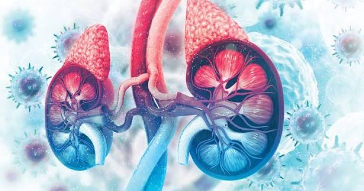
In parallel with central nervous system injury, very long-chain fatty acid accumulation inflicts severe structural damage upon the steroid-producing zones of the adrenal cortex, specifically the zona fasciculata and zona reticularis. Adrenocortical cells rely on cholesterol esters as the primary substrate for synthesizing glucocorticoids and adrenal androgens. In adrenoleukodystrophy, saturated VLCFAs preferentially esterify with cholesterol, forming insoluble cholesteryl-VLCFA crystals and lamellar lipid inclusions that are resistant to hydrolysis by neutral cholesterol ester hydrolase. As these toxic lipid aggregates accumulate inside adrenocortical cells, they trigger endoplasmic reticulum stress, mitochondrial impairment, and progressive cellular atrophy. Additionally, abnormal incorporation of VLCFAs into the plasma membrane alters membrane microviscosity, blunting the binding affinity of adrenocorticotropic hormone (ACTH) to its melanocortin-2 surface receptors. Together, cellular atrophy and receptor desensitization precipitate primary adrenocortical insufficiency (Addison’s disease), resulting in life-threatening cortisol deficiency, compensatory elevations in circulating ACTH, skin hyperpigmentation, hypoglycemia, and acute adrenal crises.
Clinical Phenotypes and Neurological Progression
Despite sharing identical ABCD1 genetic mutations—even among siblings within the same family—adrenoleukodystrophy manifests across several distinct clinical phenotypes dictated by complex environmental and modifier gene interactions. The most devastating form is Childhood Cerebral Adrenoleukodystrophy (CCALD), affecting roughly thirty-five to forty percent of boys between four and ten years of age. Previously healthy children initially exhibit subtle behavioral changes, declining academic performance, attention deficits, and emotional lability, which rapidly progress within months to cortical blindness, auditory processing loss, spastic quadriparesis, dysphagia, seizures, and a vegetative state. Conversely, Adrenomyeloneuropathy (AMN) represents the most common adult phenotype, typically emerging between the second and fourth decades of life. AMN manifests as a slowly progressive, non-inflammatory distal axonopathy affecting the dorsal columns and corticospinal tracts of the spinal cord, producing progressive spastic paraparesis, sensory ataxia, bladder dysfunction, and peripheral neuropathy. Roughly twenty percent of adult males with AMN subsequently convert to the rapidly fatal adult cerebral inflammatory phenotype.
Diagnostic Protocols: Biochemical Assays and Neuroimaging
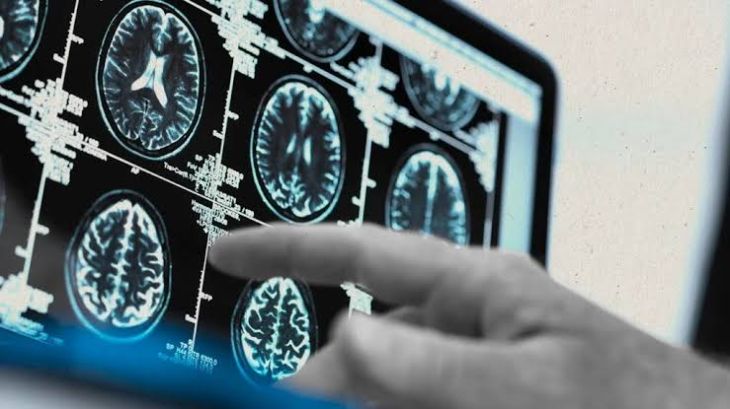
Early diagnosis of adrenoleukodystrophy—ideally through universal newborn screening prior to symptom onset—is crucial because therapeutic interventions are effective only before advanced neurological injury occurs. Biochemical diagnosis relies on quantifying plasma very long-chain fatty acid concentrations via gas chromatography-mass spectrometry (GC-MS) or liquid chromatography-tandem mass spectrometry (LC-MS/MS), demonstrating markedly elevated C26:0 levels alongside elevated C26:0/C22:0 and C24:0/C22:0 ratios. Molecular genetic sequencing of the ABCD1 gene provides definitive diagnostic confirmation and facilitates family carrier screening. In diagnosed patients, serial brain Magnetic Resonance Imaging (MRI) serves as the indispensable tool for detecting early cerebral inflammatory demyelination. Classic MRI findings demonstrate symmetric, confluent T2-weighted and FLAIR hyperintensities originating within the splenium of the corpus callosum or the pyramidal tracts and advancing outward across the parieto-occipital white matter, accompanied by a characteristic rim of gadolinium contrastenhancement marking the active inflammatory front.
Therapeutic Interventions: Transplantation, Gene Therapy, and Hormone Replacement
Management of adrenoleukodystrophy requires a dual therapeutic approach addressing both endocrine failure and neurological demyelination. Primary adrenal insufficiency is managed effectively with lifelong daily glucocorticoid replacement therapy—such as hydrocortisone or prednisone—paired with stress-dose adjustments during acute illness to prevent fatal adrenal crises, though hormone replacement does not alter neurological progression. For boys identified with early-stage cerebral adrenoleukodystrophy prior to severe cognitive or motor impairment, Allogeneic Hematopoietic Stem Cell Transplantation (HSCT) remains the established standard of care. Donor-derived myeloid precursors migrate across the blood-brain barrier and differentiate into functional microglia capable of expressing normal ALDP, arresting neuroinflammation and stabilizing white matter lesions within six to twelve months post-transplant. For patients lacking a matched donor, autologous ex vivo lentiviral Gene Therapy (elivaldogene autotemcel) transduces the patient’s own hematopoietic stem cells with functional ABCD1 complementary DNA before reinfusion, halting cerebral disease progression without graft-versus-host disease risks.
Conclusion
Adrenoleukodystrophy is a complex X-linked peroxisomal disorder driven by ABCD1 gene mutations that impair very long-chain fatty acid degradation, leading to systemic lipid toxicity across neural and endocrine tissues. Understanding how saturated VLCFA accumulation destabilizes myelin membranes, triggers microglial-driven cerebral neuroinflammation, and induces adrenocortical atrophy clarifies the clinical divergence between rapidly progressive childhood cerebral demyelination and chronic adult adrenomyeloneuropathy. Utilizing newborn tandem mass spectrometry screening, ABCD1 sequencing, and serial brain magnetic resonance imaging enables clinicians to identify cerebral inflammation during its earliest reversible window. Through timely adrenal hormone replacement alongside allogeneic stem cell transplantation or autologous lentiviral gene therapy, multidisciplinary medical teams can arrest white matter destruction and preserve neurological function for affected individuals.