What Is Rasmussen’s Encephalitis and How Can It Affect Brain Function?
Executing complex cognitive, motor, and sensory functions requires an intact, bilaterally organized cerebral cortex governed by precise neural communication and balanced metabolic activity. Rasmussen's encephalitis represents an extremely rare, severe progressive neurodegenerative disorder defined by chronic, localized inflammation restricted almost entirely to a single cerebral hemisphere. First characterized by neurosurgeon Theodore Rasmussen in the mid-twentieth century, this condition primarily afflicts young children, although adolescent and adult-onset forms are documented. The hallmark pathophysiological features of Rasmussen's encephalitis include intractable focal motor seizures, progressive hemiparesis, cognitive decline, and marked unihemispheric cerebral atrophy. Unlike multi-focal or generalized inflammatory brain disorders, Rasmussen's encephalitis selectively destroys one hemisphere while leaving the contralateral cerebral hemisphere structurally and functionally preserved during the early and intermediate stages of disease progression.
Immune-Mediated Pathophysiology and Cytotoxic Neuroinflammation
The underlying etiology of Rasmussen's encephalitis centers on a chronic, immune-mediated inflammatory response within the central nervous system. Modern neuropathological studies demonstrate that the primary drivers of tissue destruction are CD8+ cytotoxic T lymphocytes, rather than primary autoantibody deposition or active viral infection. These autoreactive CD8+ T cells infiltrate the cerebral parenchyma and directly target neurons and astrocytes. Upon recognizing unidentified neural antigens, the cytotoxic T lymphocytes release pore-forming proteins such as perforin and granzymes, inducing cell membrane damage and initiating programmed cell death (apoptosis) in affected brain cells. Microglia, the resident immune cells of the central nervous system, become intensely activated, releasing neurotoxic cytokines, reactive oxygen species, and nitric oxide. This persistent inflammatory cascade destroys synaptic connections, disrupts local blood-brain barrier integrity, and leads to extensive neuronal loss and reactive astrogliosis across the affected hemisphere.
Epileptogenesis and Mechanics of Epilepsia Partialis Continua
The progressive neuroinflammatory process severely destabilizes cortical networks, creating a highly epileptogenic environment within the involved hemisphere. As inflammatory cytokines alter neurotransmitter receptor dynamics and neuronal membrane potentials, local inhibitory networks fail, triggering continuous, uninhibited neuronal firing. A key clinical hallmark of Rasmussen's encephalitis is epilepsia partialis continua—a form of localized status epilepticus characterized by persistent, rhythmic clonic muscle twitching that affects one side of the body, most frequently involving the face, distal arm, or leg. Epilepsia partialis continua can persist continuously for hours, days, or weeks, remaining largely resistant to conventional anti-seizure medications. Over time, these continuous focal seizures spread across the entire hemisphere, evolving into complex partial seizures and secondary generalized tonic-clonic seizures, which further exacerbate metabolic strain and neuronal damage within the affected brain tissue.
Progressive Neurological Decline and Hemispheric Dysfunction
As chronic T-cell infiltration and continuous seizure activity drive unrelenting tissue destruction, patients experience progressive neurological deficits corresponding strictly to the functions managed by the damaged hemisphere. Motor deficits manifest as progressive hemiparesis, where weakness on one side of the body gradually worsens into complete hemiplegia. When the dominant cerebral hemisphere—typically the left hemisphere—is affected, patients develop severe progressive language impairment, including expressive and receptive aphasia. Parietal lobe involvement disrupts spatial orientation, sensory perception, and body schema awareness, while occipital lobe damage induces contralateral homonymous hemianopia, eliminating half of the visual field in both eyes. Concurrently, the cumulative burden of tissue loss, persistent seizure discharges, and continuous neuroinflammation leads to global cognitive decline, memory impairment, and severe behavioral alterations.
Clinical Presentation and Disease Staging Dynamics
The clinical trajectory of Rasmussen's encephalitis typically follows three distinct, sequential phases: the prodromal phase, the acute phase, and the chronic residual phase. The prodromal phase can last from several months to a few years, characterized by mild hemiparesis or low-frequency focal seizures that are often misdiagnosed as focal epilepsy or localized encephalitis. The disease then escalates into the acute phase, marked by explosive seizure frequency, the emergence of epilepsia partialis continua, rapid deterioration of motor and cognitive abilities, and accelerating hemispheric tissue loss. After several months to a year, the condition transitions into the chronic residual phase. During this stage, active neuroinflammation and seizure frequency may stabilize or diminish, but the patient is left with fixed, severe neurological deficits, including dense hemiplegia, cognitive disability, and permanent structural atrophy of the affected hemisphere.
Diagnostic Evaluation Protocols and Multimodal Neuroimaging
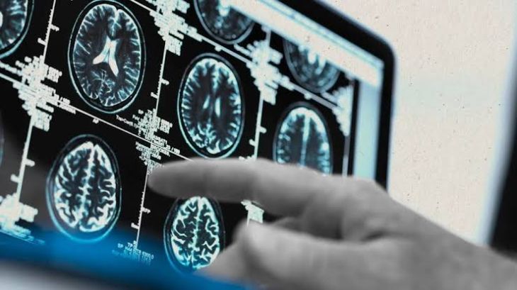
Establishing an accurate, early diagnosis of Rasmussen's encephalitis requires a comprehensive evaluation integrating clinical presentation, serial electroencephalography (EEG), advanced neuroimaging, and cerebrospinal fluid analysis. Serial Magnetic Resonance Imaging (MRI) plays a pivotal role in tracking disease progression, demonstrating early focal hyperintensities on T2-weighted and FLAIR sequences that evolve into progressive, widespread unihemispheric cortical atrophy and ex-vacuo ventricular dilation. Functional imaging using Fluorodeoxyglucose Positron Emission Tomography (FDG-PET) or Single-Photon Emission Computed Tomography (SPECT) reveals marked hemispheric hypometabolism and hypoperfusion, often preceding overt structural changes on MRI. Electroencephalography shows continuous background slowing and multifocal subcortical or cortical epileptiform discharges localized strictly to the diseased hemisphere. Brain biopsy, though rarely necessary when non-invasive criteria are met, confirms the diagnosis by showing T-cell predominant perivascular cuffing, microglial nodules, and neuronophagia.
Therapeutic Management and Surgical Intervention Strategies
Managing Rasmussen's encephalitis presents significant clinical challenges, as standard anti-seizure medications are almost universally ineffective at controlling the underlying epilepsia partialis continua or halting disease progression. Immunomodulatory therapies—including high-dose intravenous corticosteroids, plasma exchange, intravenous immunoglobulin (IVIG), and immunosuppressive drugs like tacrolimus or rituximab—are frequently deployed during the acute phase. While these medical therapies can temporarily reduce neuroinflammatory intensity and slow functional decline, they rarely prevent eventual long-term tissue destruction. Surgical intervention remains the only definitive, curative treatment for the intractable epilepsy associated with Rasmussen's encephalitis. Functional or anatomical hemispherectomy—a surgical procedure that disconnects or removes the diseased cerebral hemisphere from the rest of the brain—effectively isolates the epileptogenic tissue. When performed in young children, cerebral plasticity allows the remaining healthy hemisphere to take over many complex motor, sensory, and language functions, halting seizure activity and enabling significant cognitive recovery.
Conclusion
Rasmussen's encephalitis is a devastating, progressive neuroinflammatory disorder defined bycytotoxic T-cell-mediated destruction of a single cerebral hemisphere. Understanding how this localized inflammation drives epilepsia partialis continua, unihemispheric atrophy, progressive hemiparesis, and cognitive decline clarifies the profound functional impact experienced by affected individuals. Utilizing serial MRI, functional PET imaging, and continuous EEG monitoring allows multidisciplinary medical teams to establish an early diagnosis and differentiate Rasmussen's encephalitis from alternative focal epilepsies. Through early immunomodulatory control and timely surgical hemispherectomy, healthcare professionals can successfully eliminate intractable seizure activity, maximize neurodevelopmental plasticity, and optimize long-term quality of life for individuals living with this complex neurological condition.