What Is Posterior Cortical Atrophy and How Can It Affect Vision and Spatial Awareness?
Visual perception involves far more than simply capturing light through the ocular apparatus; it depends on an intricate, layered network of cerebral processing regions that interpret spatial positioning, physical form, motion, and depth. Posterior Cortical Atrophy represents a distinct, progressive neurodegenerative condition marked by focal, structural degeneration within the posterior cerebral cortex, particularly the occipital, parietal, and rear temporal lobes. Widely recognized as a visual or spatial presentation of Alzheimer's pathology, Posterior Cortical Atrophy manifests through early, predominant impairments in higher-order visual processing, spatial integration, and visually guided movement. Unlike typical amnestic Alzheimer's presentations where short-term memory loss dominates early stages, individuals affected by Posterior Cortical Atrophy preserve episodic memory, self-awareness, speech, and executive reasoning initially, while experiencing severe difficulties comprehending their surrounding visual environment.
Pathophysiology and Regional Neurodegenerative Patterns
On a cellular level, Posterior Cortical Atrophy is most frequently caused by abnormal neurofibrillary tau tangles and amyloid-beta plaque accumulation, matching the cellular pathology seen in standard Alzheimer's cases. Rather than concentrating within hippocampal structures and medial temporal areas, pathological aggregates predominantly target the temporoparietal and parietal-occipital cortices. Less frequently, this clinical syndrome can stem from underlying Lewy Body Dementia, Corticobasal Degeneration, or Creutzfeldt-Jakob Disease. This localized tissue degradation triggers progressive neuronal loss, synaptic breakdown, and cortical thinning along both the dorsal and ventral visual pathways. The dorsal pathway, extending toward the parietal region, governs spatial orientation and motor guidance, whereas the ventral stream, extending into temporal regions, processes object identity, facial features, and written characters.
Disruption of Spatial Processing and Balint's Syndrome
Degeneration along the dorsal visual processing stream impairs spatial orientation and undermines motor execution guided by sight. A primary clinical highlight of advanced Posterior Cortical Atrophy is Balint's syndrome, a triad composed of simultanagnosia, optic ataxia, and ocular apraxia. Simultanagnosia—the inability to perceive multiple visual elements simultaneously—prevents individuals from recognizing full scenes, turning activities like reading or moving through familiar spaces into major challenges. Optic ataxia hinders accurate reaching toward visual targets despite intact muscle strength and tactile sensitivity. Ocular apraxia limits voluntary eye movements, leaving patients unable to shift their gaze intentionally toward objects in their visual field. Together, these deficits disrupt spatial awareness, leading to frequent bumping into surroundings, miscalculated distances, and an inability to perceive environmental surroundings as a unified whole.
Ventral Stream Impairment and Agnosic Visual Deficits
As neurodegenerative changes spread into ventral occipitotemporal structures, patients develop progressive visual agnosias that disrupt object and feature recognition. Affected individuals experience prosopagnosia, rendering them unable to identify familiar faces visually and forcing reliance on voice tones or movement habits for recognition. Visual object agnosia impairs the capacity to recognize everyday items through sight alone, even while basic ocular parameters like pupil responsiveness and light reception remain normal. Additionally, alexia—severe reading difficulty—emerges rapidly, resulting from both dorsal spatial tracking errors across printed lines and ventral processing failures in recognizing letter structures and whole word forms. Patients can also develop environmental agnosia, losing their grasp of familiar landmarks, transit routes, or building interiors, which further heightens spatial confusion.
Clinical Manifestations and Impact on Daily Functionality
The gradual onset of Posterior Cortical Atrophy usually appears between fifty and sixty-five years of age, often arising earlier than traditional Alzheimer's memory symptoms. Initial complaints are frequently misattributed to eye problems, prompting repeated visits to optometrists or ophthalmologists for new glass prescriptions without success. Patients encounter difficulties reading continuous passages, managing financial figures, completing mental arithmetic (acalculia), gauging depth while driving, or dressing due to apraxia. As parietal dysfunction progresses, features of Gerstmann's syndrome—such as finger agnosia, left-right confusion, acalculia, and agraphia—may surface. Simple daily tasks like pouring a drink, arranging utensils, or spotting an item on a table become increasingly difficult, requiring substantial assistance from caregivers.
Diagnostic Evaluation Protocols and Multimodal Imaging
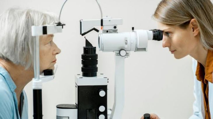
Reaching an accurate diagnosis of Posterior Cortical Atrophy requires a thorough evaluation combining specialized eye exams, formal neuropsychological testing, and advanced neuroimaging modalities. Routine ophthalmic evaluations help rule out primary ocular disorders like cataracts, glaucoma, or retinal abnormalities. Neuropsychological testing demonstrates clear visual-spatial and visual-perceptual impairments alongside relatively preserved verbal memory and general orientation. Structural Magnetic Resonance Imaging (MRI) displays focal atrophy within the bilateral parietal, occipital, and posterior temporal regions, while sparing anterior brain structures. Functional neuroimaging, such as Fluorodeoxyglucose Positron Emission Tomography (FDG-PET) or Single-Photon Emission Computed Tomography (SPECT), reveals prominent hypometabolism and reduced blood flow across posterior brain regions, offering decisive diagnostic support.
Management Strategies and Multidisciplinary Interventions
Currently, no disease-modifying or curative treatments exist specifically for Posterior Cortical Atrophy; management focuses on symptom control, environmental adjustments, and practical supportive care. Pharmacological therapies utilized in typical Alzheimer's care, such as cholinesterase inhibitors (donepezil, rivastigmine, galantamine) and N-methyl-D-aspartate receptor antagonists (memantine), are often prescribed to support neurotransmitter balance and slow general cognitive decline. Non-pharmacological approaches remain vital for preserving safety and daily function. Occupational therapy offers tailored strategies, including high-contrast household markers, uncluttered tactile environments, audiobooks, and mobility support. Specialized visual rehabilitation programs train patients to leverage preserved visual fields and adapt to spatial challenges, while caregiver support networks help families navigate the non-memory-dominant aspects of this neurodegenerative disorder.
Conclusion
Understanding how disruption across the dorsal and ventral visual streams causes spatial disorientation, simultanagnosia, optic ataxia, and agnosic visual deficits helps clarify the functional decline experienced by patients. Combining specialized neuropsychological assessments with structural MRI and functional PET imaging enables care teams to reliably distinguish Posterior Cortical Atrophy from primary ocular conditions and standard amnestic Alzheimer's disease. Through structured environmental modifications, occupational therapy, targeted cognitive support, and caregiverguidance, healthcare professionals can effectively manage symptoms, reduce accidental injury risks,and support functional independence for individuals living with this visual-spatial disorder.