What Is Polycystic Kidney Disease and How Can It Affect Kidney Function?
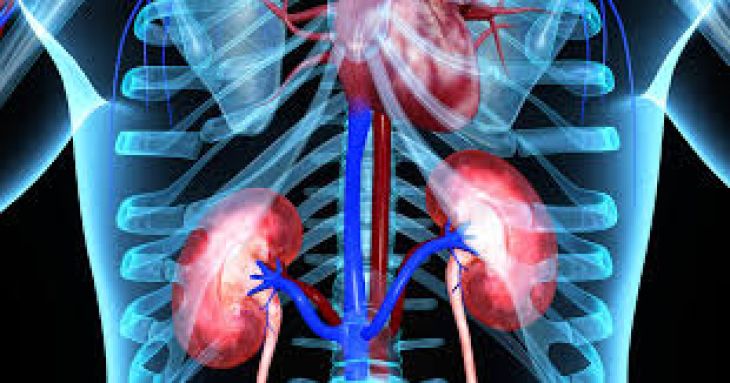
Every single day, a pair of healthy kidneys quietly processes roughly two hundred quarts of circulating blood, separating cellular waste from vital nutrients, regulating fluid volume, and keeping internal mineral levels steady. When an inherited structural disorder disrupts the physical makeup of these organs, that continuous filtration cycle encounters mounting mechanical resistance. Polycystic kidney disease—commonly abbreviated as PKD—is an inherited condition characterized by the gradual emergence and expansion of numerous fluid-filled pouches inside both kidneys. Unlike isolated, benign fluid sacs that occasionally develop during normal aging without causing harm, the growths linked to PKD multiply relentlessly and enlarge across decades. Examining the genetic origins of this disorder, its distinct inheritance patterns, and the physical ways expanding fluid sacs crowd out working tissue clarifies how PKD diminishes renal performance over time and why proactive care makes a measurable difference.
The Genetic Blueprint Behind Cyst Formation
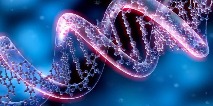
Rather than stemming from dietary habits, environmental exposure, or bacterial infections, polycystic kidney disease originates directly within a person's inherited genetic code. Most affected individuals receive an altered gene from one or both parents that governs how microscopic drainage tubes inside the kidneys are constructed and maintained. Under normal circumstances, specialized proteins act like tiny biological antennae along the inner lining of kidney tubules, sensing fluid movement and signaling cells when to stop dividing. When a genetic variation impairs those signaling proteins, isolated segments of the microscopic tubules begin ballooning outward. Over months and years, these tiny bulges pinch away from the main drainage channel, trap watery fluid inside, and continue swelling independently. Because every renal cell carries that same genetic instruction from birth, fluid sacs typically appear across both kidneys simultaneously, initiating slow anatomical changes long before any outward discomfort becomes noticeable.
Comparing the Two Inherited Forms of PKD
Medical specialists divide inherited polycystic kidney disease into two distinct categories depending on how the responsible gene travels through families and how early symptoms appear. The vastly more prevalent form passes down through a dominant inheritance pattern, representing roughly ninety percent of all diagnosed cases. Under this pattern, receiving just a single altered gene copy—most frequently involving the PKD1 or PKD2 genetic loci—from one affected parent is sufficient to trigger the disorder, giving each child a fifty percent likelihood of inheriting the trait. Individuals with this dominant form usually feel completely healthy throughout youth and early adulthood, often discovering the condition between ages thirty and fifty once enlarging sacs begin stretching the sensitive outer lining of the kidneys. Conversely, the recessive form of the disorder occurs far less frequently and follows a much more aggressive timeline. Linked to alterations in the PKHD1 gene, this rarer variant requires a child to inherit two changed gene copies—one from each healthy carrier parent. Because tubular swelling begins rapidly during fetal growth, severe kidney enlargement and accompanying liver complications often surface before birth or within the first months of life.
How Growing Fluid Sacs Crowd Out Healthy Filtration Units
Inside a normal adult kidney, roughly one million microscopic filtering clusters—known within medicine as nephrons—operate in tightly packed columns to purify blood and produce urine. As polycystic kidney disease advances, hundreds or even thousands of liquid-filled pockets form throughout both organs, ranging in diameter from tiny pinpoint bubbles to spheres rivaling a grapefruit. Crucially, this dramatic growth adds zero filtering capability; instead, the swelling fluid pockets press firmly against neighboring nephrons, pinch tiny surrounding blood vessels, and provoke localized tissue inflammation that gradually hardens into non-functional scar tissue. As working filtration clusters get squeezed out and replaced by fluid cavities and fibrous scarring, the organs progressively lose their capacity to clear metabolic toxins from the bloodstream.
Early Physical Clues and Beyond-the-Kidney Effects
Because undamaged nephrons initially work harder to pick up the slack for compressed neighbors, many adults live with polycystic kidney disease for decades without suspecting an underlying issue. Frequently, the very first clinical warning sign is elevated blood pressure, which arises when expanding cysts squeeze internal renal arteries and prompt the kidneys to release blood-vessel-constricting hormones into circulation. As overall kidney volume increases, people commonly notice a persistent, dull ache or feeling of fullness across the lower back, flanks, or abdomen caused by tension on the organ's outer fibrous covering. Occasional pink or reddish urine may appear when fragile surface vessels along a cyst wall break open, while sluggish urine drainage inside distorted kidneys creates favorable conditions for urinary infections and mineral stones. Additionally, because the underlying genetic variations influence structural proteins in other tissues, some individuals also develop harmless fluid sacs in the liver, floppy heart valves, small outpouchings along the intestinal wall, or localized weak spots in cranial blood vessels that require periodic monitoring.
Gradual Progression Toward Reduced Kidney Capacity
Across several decades, the steady expansion of fluid pockets and surrounding scar tissue slowly lowers the speed at which the kidneys filter waste—a clinical measurement tracked through routine blood tests. Once overall filtration efficiency falls consistently below healthy benchmarks, the condition enters the spectrum of ongoing kidney impairment. Metabolic byproducts such as urea and creatinine start lingering in the bloodstream, excess fluid may accumulate around the ankles and lower legs, and reduced kidney hormone output can contribute to low red blood cell counts and diminished bone density. When functional tissue drops below a critical threshold, the kidneys can no longer maintain internal chemical balanceon their own, making regular blood-filtering dialysis treatments or a donor kidney transplant necessary to sustain health. Notably, the timeline of this decline differs considerably from one person to another; individuals carrying PKD2 genetic variations frequently preserve reliable filtration wellinto their sixties or seventies, whereas certain PKD1 variations tend to drive faster cyst enlargement earlier in adulthood.
Diagnostic Evaluation and Strategies to Protect Kidney Health
Identifying polycystic kidney disease generally starts with non-invasive imaging alongside a detailed review of family health history. Abdominal ultrasound serves as the primary screening tool, using reflected sound waves to count and measure fluid-filled sacs across both kidneys, while magnetic resonance or computed tomography scans calculate total kidney volume with high precision to monitor growth rates over time. While current therapies cannot alter the inherited DNA instructions that spark cyst formation, consistent medical care can meaningfully slow functional decline and safeguard heart health. Keeping blood pressure tightly controlled—typically using targeted blood pressure medications that relax renal blood vessels—reduces physical strain on remaining nephrons. For adults showing signs of rapid kidney enlargement, specialized prescription therapies can interrupt cellular signals that pump fluid into growing sacs. Pairing medical treatment with generous daily water consumption, lower dietary sodium, consistent aerobic exercise, and careful avoidance of kidney-irritating over-the-counter pain relievers helps preserve working kidney tissue for as many years as possible.
Conclusion
Polycystic kidney disease represents an inherited structural challenge that reshapes the kidneys slowly through the relentless growth of fluid-filled sacs. By compressing delicate filtering units, narrowing internal blood vessels, and encouraging scar tissue buildup, these expanding pockets steadily reduce the organs' vital ability to clear waste, regulate body fluids, and stabilize blood pressure. Recognizing early indicators such as unexplained high blood pressure or persistent side discomfort enables timely diagnosis before widespread tissue loss occurs. Through regular imaging checkups, disciplined blood pressure management, targeted medications, and kidney-protective lifestyle choices, individuals living with PKD can actively defend their remaining renal function and enjoy full, active lives for decades.