What Is Hypoplastic Left Heart Syndrome and How Does It Affect Heart Function?
In a fully formed human heart, two distinct pumping sides work in continuous rhythm—the right side sends oxygen-poor blood toward the lungs, while the muscular left side propels oxygen-rich blood out to nourish the brain, organs, and limbs. When structural development stalls during the earliest weeks of pregnancy, however, a baby can enter the world with a critically incomplete circulatory engine. Hypoplastic left heart syndrome, frequently shortened to HLHS, is a complex congenital heart defect in which the structures along the left side of the heart fail to grow to their normal size and strength before birth. Because the left ventricle, its associated valves, and the main outgoing artery remain severely undersized, the heart cannot pump oxygenated blood throughout the body on its own. Exploring the anatomical abnormalities behind this condition, how circulation shifts immediately after delivery, and the specialized surgical stages used to rebuild blood flow reveals how HLHS alters cardiac performance and how modern pediatric heart care sustains life.
Anatomical Structures Affected on the Left Side of the Heart
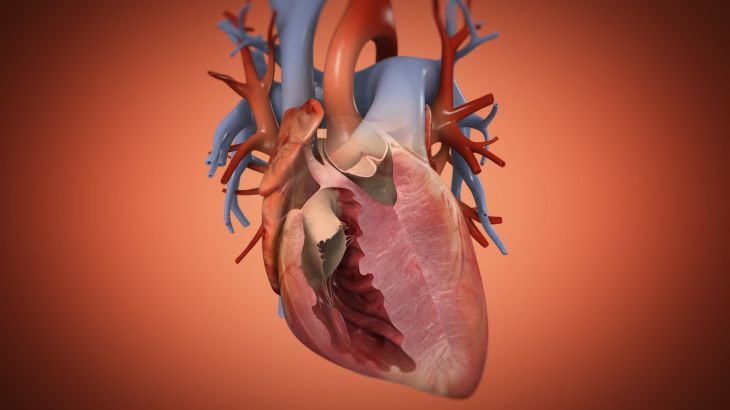
To grasp how hypoplastic left heart syndrome disrupts circulation, it helps to examine the specific chambers and pathways that fail to mature properly during fetal growth. In a baby born with HLHS, the lower left pumping chamber—known as the left ventricle—is strikingly small and thin-walled, lacking the muscular force needed to push blood into systemic circulation. Alongside this underdeveloped chamber, the mitral valve, which normally regulates blood flow between the upper and lower left chambers, may be unusually narrow or completely sealed shut. Similarly, the aortic valve leading out of the left ventricle is often severely constricted or fused, preventing blood from exiting forward. Beyond the heart chambers themselves, the ascending aorta—the main arterial trunk responsible for distributing oxygen-rich blood across the entire body—is typically thread-thin and underdeveloped, leaving the left side of the heart anatomically unable to fulfill its primary workload.
Why Circulation Changes Dramatically Right After Birth
During pregnancy, a fetus with hypoplastic left heart syndrome usually grows comfortably inside the womb because the mother's placenta handles oxygen exchange and two temporary fetal pathways keep blood moving around the underdeveloped left chambers. The first temporary bridge, called the foramen ovale, is a small natural opening between the upper right and left collecting chambers, while the second, known as the ductus arteriosus, connects the lung artery directly to the body's main artery. Before birth, the stronger right side of the heart uses these two shortcuts to supply blood to both the developing lungs and the rest of the fetal body. Once the baby takes its first breaths after delivery, however, normal biological signals trigger both temporary pathways to seal shut within hours or days. As the ductus arteriosus narrows and closes, blood flow to the brain, kidneys, and extremities abruptly drops, plunging the newborn into a life-threatening circulatory crisis unless immediate medical intervention keeps those fetal channels open.
How Single-Ventricle Strain Alters Overall Heart Performance
With the left side unable to contribute meaningful pumping power, the entire burden of sustaining circulation falls onto the right ventricle—a chamber naturally built to push blood through the low-resistance blood vessels of the lungs rather than the high-pressure arterial network of the whole body. In newborns with hypoplastic left heart syndrome, oxygen-rich blood returning from the lungs must cross over through an opening between the upper chambers into the right side, where it mixes with oxygen-poor blood returning from the body. The right ventricle then pumps this blended, partially oxygenated blood out toward both the lungs and the systemic arteries simultaneously. Because a single chamber is performing double duty against much higher resistance than intended, the heart beats faster, works under elevated wall stress, and delivers lower overall oxygen saturation to vital tissues—a combination that rapidly exhausts the infant without targeted stabilization.
Early Warning Signs and Prenatal or Newborn Detection
Many cases of hypoplastic left heart syndrome are identified before delivery during routine mid-pregnancy anatomy ultrasounds, where specialized imaging highlights an asymmetric four-chamber heart view. When the condition goes undetected prior to birth, infants often appear healthy during their very first hours while the temporary fetal vessels remain open. As those pathways begin closing over the first few days of life, telltale signs of failing systemic blood flow emerge rapidly. Parents and neonatal nurses may notice a bluish or grayish tint around the lips, tongue, and fingernails caused by reduced oxygen levels, accompanied by rapid, labored breathing and flaring nostrils. The newborn may become unusually lethargic, struggle to feed without tiring, develop cold hands and feet, or show weak pulses in the legs and arms—all urgent clinical signals that systemic circulation is faltering.
The Three-Stage Surgical Reconstruction Pathway
Because medicine cannot regrow a missing left ventricle, pediatric heart surgeons treat hypoplastic left heart syndrome by re-engineering the circulatory system through a staged series of three open-heart procedures so the right ventricle can reliably serve as the body's main pump. Within the first week of life, surgeons perform the Norwood procedure, rebuilding the narrow aorta using tissue from the pulmonary artery and placing a small synthetic tube to maintain steady blood flow to the lungs. Between four and six months of age, the second stage—known as the Glenn procedure—reroutes oxygen-poor blood returning from the upper body directly into the lung arteries, relieving half of the volume workload from the single pumping ventricle. Finally, between eighteen months and four years of age, the Fontan procedure directs oxygen-poor blood from the lower half of the body straight into the lungs as well. Once all three stages are complete, oxygenated and deoxygenated blood no longer mix, allowing the right ventricle to focus exclusively on pumping oxygen-rich blood to the body.
Long-Term Outlook and Ongoing Cardiac Care
While staged surgical reconstruction has transformed hypoplastic left heart syndrome from a universally fatal diagnosis into a survivable condition, living with a single-ventricle circulation requires lifelong partnership with specialized congenital heart teams. Because the right ventricle is anatomically shaped differently than a true left ventricle, it remains vulnerable over the decades to gradual muscle fatigue, leaky heart valves, irregular heart rhythms, and elevated pressure inside the lung vessels and liver. Regular echocardiograms, cardiac magnetic resonance imaging, exercise capacity assessments, and tailored medications help doctors spot subtle changes in pumping efficiency early. Many children and young adults who undergo successful staged reconstruction attend school, participate in approved recreational activities, and lead fulfilling lives, though some may eventually require catheter-based interventions or heart transplantation later in adulthood if the single pumping chamber begins to tire.
Conclusion
Hypoplastic left heart syndrome profoundly reshapes cardiac function by leaving the left side of the heart too small to circulate oxygenated blood, forcing the right ventricle to shoulder the entire circulatory workload alone. Although temporary fetal blood vessels briefly mask the defect immediately after birth, their natural closure triggers an urgent need for medication and staged surgical reconstruction. Through the coordinated sequence of the Norwood, Glenn, and Fontan operations, surgeons separate oxygen-rich and oxygen-poor blood pathways, dramatically improving oxygen delivery and survival. Combined with attentive lifelong monitoring from congenital cardiology specialists, early diagnosis and modern surgical care give children born with HLHS the opportunity to grow, thrive, and navigate life with resilient heart function.