What Is a Coronary Artery Fistula and How Can It Affect Blood Flow?
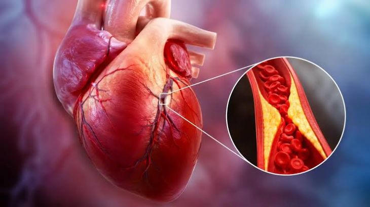
Maintaining adequate myocardial perfusion requires an intact coronary arterial network capable of delivering oxygenated blood under high pressure directly to the capillary beds of the ventricular myocardium. A coronary artery fistula represents an uncommon congenital or acquired vascular anomaly characterized by an abnormal direct communication between a coronary artery and a low-pressure cardiac chamber, systemic vein, or pulmonary vessel, bypassing the normal myocardial capillary bed. Under normal anatomical conditions, the right and left coronary arteries arise from the aortic sinuses and branch progressively into smaller arterioles to supply the surrounding muscular tissue. When a fistula forms, it creates an anatomical shunt that diverts blood flow away from high-resistance myocardial capillaries into lower-resistance vascular spaces or heart chambers. This structural abnormality disrupts normal coronary hemodynamics, establishing an altered pressure gradient that can impair myocardial perfusion, provoke tissue ischemia, and induce progressive cardiac remodeling.
Etiology, Embryological Origins, and Structural Characteristics
The underlying origin of a coronary artery fistula can be categorized into congenital developmental errors or acquired vascular disruptions. Congenital fistulas account for the vast majority of cases, resulting from incomplete involution of the embryonic intramyocardial trabecular space or abnormal persistence of primitive sinusoidal networks during early cardiac development. In healthy embryonic growth, these sinusoidal spaces gradually compress into normal capillary networks; failure of this regression leaves persistent, large-caliber conduits between major coronary arteries and internal cardiac structures. Acquired fistulas develop secondary to blunt or penetrating chest trauma, iatrogenic injury during invasive cardiac procedures such as endomyocardial biopsy, pacemaker lead placement, or coronary bypass grafting, or as a complication of severe coronary vasculitis. Congenitally, the right coronary artery or its branches serve as the origin site in more than half of reported cases, followed by the left anterior descending artery, with the majority of fistulas terminating into right-sided, low-pressure structures like the right ventricle, right atrium, or pulmonary artery.
Mechanics of Coronary Steal and Hemodynamic Impairment
The primary pathophysiological mechanism through which a coronary artery fistula causes myocardial ischemia is the phenomenon known as coronary steal syndrome. Because fluid naturally follows the path of least resistance, blood flowing through the feeding coronary artery preferentially drains into the low-pressure recipient chamber or vessel rather than entering the high-resistance capillary bed of the distal myocardium. During periods of resting physiological state, baseline blood flow may remain adequate to meet metabolic demands. However, during physical exertion or stress, when myocardial oxygen demand increases dramatically, the resistance in the fistulous tract remains significantly lower than that of the dilated downstream microvascular bed. As a result, oxygenated blood is diverted or "stolen" away from the myocardium distal to the fistula site, producing localized myocardial ischemia despite the absence of atherosclerotic plaque obstruction.
Volumetric Overload, Chamber Dilation, and Long-Term Complications
In addition to localized perfusion deficits, a coronary artery fistula imposes a chronic volumetric strain on both the coronary arteries and the recipient cardiac chambers. The continuous, high-volume flow traversing the feeding coronary artery subjects its vascular wall to elevated shear stress, triggering compensatory arterial dilation, vascular tortuosity, and eventual aneurysmal formation. Over time, these dilated arterial segments become prone to mural thrombus formation, spontaneous dissection, or distal thromboembolism. Furthermore, the continuous shunting of blood alters cardiac chamber volume dynamics. When the fistula drains into right-sided cardiac structures, it creates a left-to-right shunt, increasing pulmonary blood flow and inducing chronic volume overload on the right atrium and right ventricle. Over prolonged periods, this continuous volumetric strain can promote progressive pulmonary arterial hypertension, right ventricular hypertrophy, structural heart failure, and atrial or ventricular dysrhythmias.
Clinical Presentation and Diagnostic Evaluation Protocols
The clinical spectrum of coronary artery fistulas ranges from completely asymptomatic, incidentally discovered structural anomalies to overt exertional angina, heart failure, or sudden cardiac collapse. Asymptomatic presentations are common in pediatric populations or individuals with small-caliber shunts. However, as age advances or as the fistulous tract enlarges, patients frequently develop exertional dyspnea, chest pain, exercise intolerance, fatigue, and palpitations. Physical examination often reveals a continuous, machine-like murmur heard best over the lower sternal border, reflecting persistent blood flow throughout both systole and diastole. Diagnosing a coronary artery fistula requires detailed multimodality cardiac imaging. Transthoracic and transesophageal echocardiography can identify dilated coronary origins, abnormal turbulent color flow jets, and enlarged recipient chambers. Invasive coronary angiography remains the definitive gold standard, precisely mapping the origin, course, caliber, and insertion site of the fistulous tract, while multi-detector Computed Tomography (CT) angiography provides comprehensive three-dimensional anatomical visualization of surrounding structures.
Management Strategies and Intervention Protocols
Managing a coronary artery fistula depends primarily on the size of the shunt, the presence of clinical symptoms, the severity of myocardial ischemia, and the risk of long-term structural complications. Small, asymptomatic fistulas with negligible shunt fractions generally do not require immediate invasive intervention; instead, they are managed conservatively with periodic echocardiographic monitoring and clinical follow-up. Conversely, large, highly symptomatic fistulas, or those associated with significant coronary steal, progressive chamber enlargement, or high-volume shunting, warrant definitive closure to restore normal myocardial perfusion and prevent irreversible structural remodeling. Intervention options include catheter-based transcatheter embolization using vascular occlusion coils, covered stents, or vascular plugs, which offers a minimally invasive approach with favorable recovery profiles. Surgical ligation, performed with or without cardiopulmonary bypass, is reserved for complex, tortuous, or aneurysmal fistulas, multiple feeding branches, or cases requiring concurrent cardiac surgical procedures.
Conclusion
A coronary artery fistula is a distinct, uncommon vascular anomaly that disrupts normal myocardial blood flow by creating a low-resistance shunt between a coronary artery and a recipient chamber or vessel. By understanding how this abnormal communication induces coronary steal dynamics, volume overload, arterial dilation, and localized myocardial ischemia, clinicians can better comprehend the functional decline and symptom presentation observed in affected individuals. Utilizing advancednon-invasive imaging alongside invasive coronary angiography enables care teams to accurately characterize the fistula's structural trajectory and assess its hemodynamic impact. Through careful risk stratification, conservative clinical monitoring, and timely deployment of transcatheter occlusion or surgical ligation, healthcare professionals can successfully restore physiological coronary perfusion, prevent long-term cardiac complications, and safeguard patient cardiovascular health.