What Is Constrictive Pericarditis and How Does It Affect Heart Function?
Introduction to Pericardial Architecture and Structural Constriction
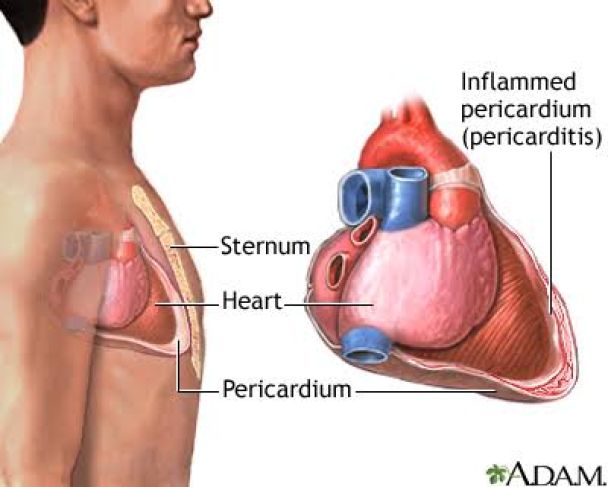
Optimizing systemic perfusion requires unimpeded ventricular relaxation throughout diastole to secure adequate stroke volumes. Constrictive pericarditis is an advanced cardiovascular disorder characterized by chronic inflammation, extensive fibrotic deposition, and frequent dystrophic calcification of the pericardial sac surrounding the myocardium. In healthy anatomy, the pericardium functions as an elastic, dual-layered sheath with minimal serous fluid that reduces friction during cardiac motion while preventing acute chamber dilatation. In constrictive disease, this flexible structure degrades into a rigid, inelastic container. This firm encasement physically restricts cardiac expansion during diastolic filling, impeding systemic venous return and disrupting global intracardiac flow dynamics.
Etiology and Pathophysiological Fibrotic Remodeling
The pathological sequence culminating in constrictive pericarditis typically originates with persistent or recurrent inflammation of the pericardial layers. In industrialized regions, idiopathic and post-viral etiologies predominate, followed by previous open-heart surgery, mediastinal irradiation, systemic autoimmune disorders, and purulent pericardial infections. The cellular mechanism entails an aberrant repair cascade in which fibrin-rich exudates undergo organization into dense fibrous tissue. Over time, sustained fibroblast activation, heavy extracellular matrix turnover, and secondary calcification fuse the visceral and parietal layers into an unyielding shell that isolates the heart from normal intrathoracic pressure swings.
Hemodynamic Alterations and Enhanced Ventricular Interdependence
The physiological hallmark of constrictive pericarditis is severe restriction of ventricular diastolic filling operating alongside heightened ventricular interdependence. Because the encased pericardium establishes a rigid maximum boundary for total cardiac volume, volume expansion in one chamber directly restricts the available space for the adjacent chamber. During inspiration, falling intrathoracic pressure augments venous flow into the right atrium and ventricle. Lacking outward expansion capacity against the rigid shell, the right ventricular free wall forces the interventricular septum to bulge leftward into the left ventricular lumen. This mechanical displacement reduces left ventricular volume and diminishes systemic stroke volume during inspiration—a state termed enhanced ventricular interdependence. Additionally, early diastolic filling proceeds rapidly until the myocardium abruptly strikes the non-compliant wall, yielding the characteristic "dip-and-plateau" configuration on pressure tracings.
Systemic Congestive Manifestations and Clinical Signs
Because ventricular relaxation is severely restricted, systemic venous pressures increase dramatically to force blood into the non-compliant heart, driving extensive backward congestive failure. Affected individuals typically exhibit an insidious onset of symptoms linked to elevated systemic venous pressures and reduced cardiac output. Elevated venous pressure presents as pronounced jugular venous distension, congestive hepatomegaly, abdominal ascites, and dependent peripheral edema. A key physical sign is Kussmaul sign, characterized by a paradoxical elevation in jugular venous pressure during inspiration because the rigid right heart cannot accommodate the inspiratory increase in venous return. Symptoms of compromised cardiac output include exertional breathlessness, chronic fatigue, severe exercise limitation, and cardiac cachexia. Cardiac auscultation often reveals a pericardial knock, an early diastolic sound caused by the sudden halting of ventricular expansion against the stiff pericardial wall.
Diagnostic Modalities and Invasive Assessment
Differentiating constrictive pericarditis from primary restrictive cardiomyopathy is a crucial diagnostic step, as both conditions present with severe diastolic filling impairment. Accurate distinction requires multimodality cardiac imaging. Transthoracic echocardiography highlights key diagnostic markers, such as respiratory variation in AV valve inflow velocities, expiratory hepatic vein flow reversal, and dynamic septal bounce. Cross-sectional modalities like Computed Tomography (CT) and Magnetic Resonance Imaging (MRI) provide precise anatomical characterization by quantifying pericardial thickness and mapping focal or diffuse calcifications. Diagnostic cardiac catheterization remains the gold standard, demonstrating end-diastolic pressure equalization across all four cardiac chambers, prominent "a" and "v" waves with sharp "x" and "y" descents on atrial tracings, and respiratory discordance between peak right and left ventricular pressures.
Management Protocols and Surgical Pericardiectomy
Treatment strategies are dictated by disease duration and the degree of structural remodeling. For transient or subacute presentations driven by ongoing active inflammation, pharmacotherapy using non-steroidal anti-inflammatory agents, colchicine, or systemic corticosteroids can successfully reduce pericardial edema and halt progression to permanent restriction. Conversely, once irreversible fibrosis and calcification establish chronic mechanical constriction, conservative medical therapy is restricted to cautious diuretic administration to ease congestive symptoms without inducing severe hypotension. Complete surgical pericardiectomy represents the definitive definitive curative intervention for established chronic constriction. This major procedure requires radical excision of the scarred, adherent pericardium between both phrenic nerves, freeing the underlying myocardium to restore normal filling dynamics and eliminate systemic venous congestion.
Conclusion
Constrictive pericarditis is a severe cardiovascular disorder where chronic inflammatory scarring replaces the elastic pericardial sac with an unyielding shell. Understanding how pericardial thickening restricts diastolic expansion, equalizes chamber pressures, and amplifies ventricular interdependence clarifies the fundamental mechanisms driving systemic venous congestion and functional decline. Combining cross-sectional imaging with invasive pressure recordings allows clinicians to confidently distinguish pericardial constriction from primary myocardial disease. Prompt surgical pericardiectomy effectively removes mechanical restriction, restores physiological diastolic filling, and alleviates chronic congestion to preserve long-term cardiac performance.