What Is Aortic Coarctation and How Does It Affect Blood Flow?
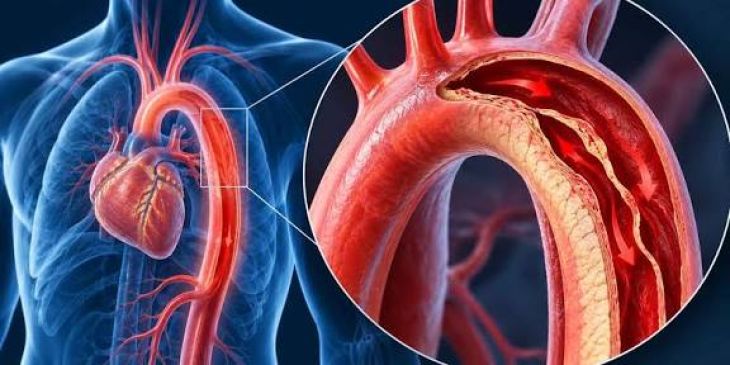
The aorta serves as the primary conduit for oxygenated blood leaving the left ventricle, distributing high-pressure systemic blood flow through its ascending, arch, thoracic, and abdominal segments. Aortic coarctation represents a major congenital cardiovascular malformation characterized by a discrete, localized narrowing of the aortic lumen. Most commonly situated in the juxtaductal position—just distal to the origin of the left subclavian artery and adjacent to the insertion of the ductus arteriosus—this anatomical constriction creates a high-resistance obstacle to systemic blood flow. By inserting a mechanical barrier within the high-volume thoracic aorta, aortic coarctation creates a profound pressure gradient between the upper body circulation proximal to the stenosis and the lower body circulation distal to it. This alteration in hemodynamics imposes a chronic pressure overload on the left ventricle while impairing perfusion to abdominal organs and lower extremities.
Etiology, Embryological Origins, and Structural Pathomorphology
The precise developmental mechanism producing aortic coarctation remains centered on two primary embryological theories: the ectopic ductal tissue theory and the hemodynamic flow theory. The ductal tissue theory posits that smooth muscle tissue from the embryonic ductus arteriosus aberrantly extends into the adjacent aortic wall during fetal development. When the ductus closes shortly after birth in response to rising arterial oxygen tension, this ectopic ductal tissue constricts as well, producing a localized shelf-like infolding of the aortic media. The hemodynamic flow theory suggests that reduced blood flow through the fetal left heart and ascending aorta—often due to associated lesions like a bicuspid aortic valve or ventricular septal defect—leads to underdeveloped aortic arch growth and localized luminal narrowing. Pathologically, the coarctation site features medial thickening, elastic fiber fragmentation, and intimate hyperplasia, rendering the localized vessel segment rigid and non-compliant.
Mechanics of Pressure Overload and Collateral Vessel Development
The mechanical constriction imposed by aortic coarctation fundamentally alters systemic arterial pressure dynamics. Proximal to the coarctation, systemic vascular resistance increases significantly, elevating systolic blood pressure within the arch arteries that supply the head, neck, and upper limbs. This severe upper-body arterial hypertension forces the left ventricle to generate markedly elevated intraventricular pressures during systole to maintain cardiac output, inducing progressive concentric left ventricular hypertrophy, increased myocardial wall stress, and elevated oxygen consumption. Conversely, distal to the coarctation, arterial blood pressure is significantly reduced, resulting in blunted, delayed arterial pulses and lower baseline perfusion pressures within the abdominal viscera and lower extremities. To bypass the narrowed aortic segment, the body develops an extensive network of collateral blood vessels, primary involving the internal thoracic, intercostal, subclavian, and scapular arteries, which dilate and hypertrophy over time to channel blood to the lower aorta.
Clinical Presentation and Upper-to-Lower Hemodynamic Dissociation
The clinical presentation of aortic coarctation varies across a broad spectrum depending on the severity of luminal narrowing and the rate of ductal closure. In severe infant presentations, rapid postnatal closure of the ductus arteriosus causes a sudden drop in lower-body perfusion, precipitating acute left heart failure, cardiogenic shock, metabolic acidosis, and oliguria. In older children and adults, the condition often remains clinically silent for years, protected by gradual collateral vessel formation. When detected, the clinical hallmark of aortic coarctation is a prominent pulse differential and blood pressure gradient between the upper and lower extremities. Physical examination typically reveals strong, bounding radial pulses alongside weak, delayed femoral pulses (radio-femoral delay), accompanied by significantly higher systolic blood pressure readings in the arms compared to the legs. A continuous or systolic ejection murmur is often heard over the left infraclavicular area and back, produced by turbulent blood flow across the narrowed segment and dilated intercostal collaterals.
Secondary Cardiovascular Complications and End-Organ Effects
Uncorrected or long-standing aortic coarctation leads to widespread systemic complications secondary to chronic upper-body hypertension and abnormal wall shear stress. Continuous elevation of proximal aortic pressure accelerates premature coronary artery disease, aortic root dilation, and aneurysm formation within the Circle of Willis, predisposing patients to stroke or fatal subarachnoid hemorrhage. The mechanical stress of turbulent blood flow immediately distal to the coarctation site frequently causes post-stenotic aortic dilation, which carries an increased long-term risk of aortic wall dissection or rupture. Furthermore, chronic lower-body hypoperfusion can trigger renal hypoperfusion signaling, activating the renin-angiotensin-aldosterone system. This endocrine response worsens systemic hypertension, further compounding left ventricular strain, heart failure risk, and hypertensive nephrosclerosis over time.
Diagnostic Protocols and Multimodal Imaging Assessment
Diagnosing aortic coarctation involves a systematic evaluation integrating physical exam findings with specialized cardiovascular imaging. Transthoracic echocardiography serves as the initial diagnostic tool, providing direct visualization of the coarctation site, measuring continuous wave Doppler peak pressure gradients, assessing left ventricular hypertrophy, and evaluating associated cardiac defects such as bicuspid aortic valves. In older patients or complex anatomies, Computed Tomographic (CT) Angiography and Cardiac Magnetic Resonance (CMR) imaging offer comprehensive three-dimensional visualization of the thoracic aorta. These advanced imaging modalities precisely quantify the minimum luminal diameter, assess collateral vessel development, measure post-stenotic dilation, and evaluate retrosternal surgical access. Invasive cardiac catheterization is reserved for cases requiring precise invasive pressure gradient measurements or simultaneous therapeutic endovascular interventions.
Therapeutic Interventions and Long-Term Management
Treatment for aortic coarctation aims to relieve the luminal obstruction, eliminate the peak pressure gradient, restore normal lower-body perfusion, and alleviate proximal hypertension. In neonates with critical coarctation, continuous intravenous prostaglandin E1 infusion is life-saving, maintaining ductal patency to preserve lower-body blood flow until definitive repair can be performed. In older infants, children, and young adults, primary therapeutic options include surgical resection or endovascular intervention. Surgical techniques—such as extended end-to-end anastomosis, subclavian flap aortoplasty, or prosthetic patch grafting—are widely utilized with high success rates. For older children and adult patients, transcatheter endovascular repair using balloon angioplasty with covered or bare-metal stent implantation has become a primary treatment modality, effectively dilating the narrowed lumen and reinforcingthe aortic wall against recoiling or aneurysm formation. Continuous long-term follow-up remains necessary to monitor for residual or recurrent hypertension, re-coarctation, and late aneurysm development.
Conclusion
Aortic coarctation is a critical congenital vascular anomaly defined by localized narrowing of the thoracic aorta, producing severe pressure dissociation between upper and lower systemic circulations. Understanding how this mechanical obstacle increases left ventricular afterload, triggers extensive collateral vessel recruitment, and causes severe proximal hypertension clarifies the cardiovascular risks and functional decline experienced by affected individuals. Utilizing detailed echocardiography, CT angiography, and cardiac MRI enables multidisciplinary clinical teams to accurately characterize the anatomical defect and quantify hemodynamic severity. Through timely surgical resection, balloon angioplasty, or endovascular stent placement, healthcare professionals can successfully eliminate the aortic gradient, restore physiological systemic blood flow, and protect long-term cardiovascular health.