Understanding Pericardial Effusion and Its Effects on the Heart
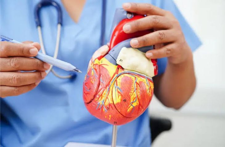
The human heart is securely suspended within the mediastinum by a specialized, double-walled fibroserous sac known as the pericardium. This anatomical housing consists of a tough, outer fibrous layer that anchors the heart to surrounding mediastinal structures and a smooth, inner serous layer divided into parietal and visceral components. Under normal physiological conditions, the potential space between these layers—known as the pericardial cavity—contains only a modest amount of lubricating serous fluid, typically measuring between fifteen and fifty milliliters. This minimal fluid volume reduces friction, allowing the heart to expand and contract smoothly with every beat throughout the cardiac cycle. However, when systemic inflammation, malignant infiltration, metabolic disorders, or traumatic injury disrupt normal fluid balance, excess fluid can accumulate rapidly or insidiously within this closed compartment, precipitating a medical condition known as pericardial effusion. Understanding the delicate pressures within the pericardial sac and the resulting impact on cardiac hemodynamics is essential for recognizing and managing this potentially life-threatening condition.
Pathological Causes and Etiologies of Pericardial Fluid Accumulation
Pericardial effusion is rarely a primary, isolated disorder; rather, it manifests as a secondary consequence of a diverse array of systemic, infectious, and inflammatory pathologies. One of the most frequent causes is acute pericarditis, an inflammation of the pericardial membranes triggered by viral infections, bacterial invasion, or post-myocardial infarction immune responses known as Dressler's syndrome. When the pericardium becomes inflamed, its capillaries leak protein-rich fluid into the pericardial space. Another major etiology is malignant disease, where metastatic cancers—most commonly lung cancer, breast cancer, or lymphoma—invade the pericardial tissue or obstruct lymphatic drainage, causing persistent, high-volume fluid accumulation. Furthermore, systemic medical conditions such as severe chronic renal failure with uremia, advanced hypothyroidism, autoimmune disorders like systemic lupus erythematosus, and physical trauma or aortic dissections can all trigger varying degrees of pericardial effusion.
The Biomechanics of the Pericardial Sac and Pressure Elevation
To comprehend how a pericardial effusion alters cardiac function, one must examine the physical properties of the pericardium itself. The outer fibrous pericardium is remarkably tough and inelastic, designed to prevent acute over-dilation of the heart during strenuous exertion. Because this sac cannot stretch rapidly when fluid accumulates slowly over weeks or months, it accommodates small increases in volume initially without causing significant pressure changes. However, once a critical volume threshold is reached, or if fluid accumulates rapidly due to trauma or hemorrhage, the compliance limits of the pericardial sac are abruptly exhausted. At this juncture, any additional drop of fluid causes intra-pericardial pressure to skyrocket, transforming the pericardial space from a compliant lubricating layer into a rigid, pressurized vise that begins to compress the thin-walled chambers of the heart.
Hemodynamic Impact and the Pathophysiology of Cardiac Tamponade
When rising fluid pressure within the pericardial sac exceeds the normal diastolic pressures inside the cardiac chambers, the heart can no longer fill properly during diastole, precipitating a catastrophic medical emergency known as cardiac tamponade. Because the low-pressure right atrium and right ventricle have thinner walls than the muscular left side of the heart, they are the first chambers to collapse under external fluid pressure. As diastolic filling of the right side of the heart becomes severely impaired, venous return from the body is obstructed, leading to a marked elevation in jugular venous pressure. Simultaneously, the inability of the ventricles to fill adequately causes a precipitous drop in stroke volume and cardiac output. This circulatory failure manifests clinically through Beck's triad—hypotension, muffled heart sounds, and distended jugular veins—alongside a phenomenon known as pulsus paradoxus, where systolic blood pressure drops abnormally during inspiration due to shifting interventricular pressures.
Clinical Presentation and Diagnostic Challenges
The clinical presentation of a pericardial effusion varies dramatically depending on the speed at which the fluid accumulates and whether it has progressed to frank cardiac tamponade. When an effusion develops slowly over a long period, the pericardium stretches gradually, allowing patients to remain remarkably asymptomatic for months, with the condition occasionally discovered incidentally during imaging studies. However, when symptoms do arise, patients commonly report a dull, oppressive chest ache, dyspnea, orthopnea, and a sense of fullness in the chest. As cardiac output declines, individuals may experience profound fatigue, lightheadedness, syncope, and cool, clammy extremities. Because these symptoms heavily overlap with other cardiopulmonary conditions like congestive heart failure or pneumonia, maintaining clinical vigilance is critical for early detection.
Diagnostic Investigations: Echocardiography and Imaging Modalities
Confirming the presence of a pericardial effusion and evaluating its hemodynamic significance relies heavily upon advanced cardiac imaging techniques.
-
Echocardiography: Transthoracic echocardiography serves as the undisputed gold standard for diagnosing pericardial effusion. Ultrasound imaging allows clinicians to visualize the fluid-filled pericardial space directly, appearing as an echo-free dark space surrounding the heart muscle. Furthermore, echocardiography provides real-time evidence of early cardiac tamponade by revealing diastolic collapse of the right atrium and right ventricle, excessive swinging motion of the heart within the fluid, and exaggerated respiratory variations in blood flow velocities across heart valves.
-
Complementary Imaging: In complex cases, chest radiography may reveal an enlarged, water-bottle-shaped cardiac silhouette, while computed tomography scans and magnetic resonance imaging provide detailed anatomical mapping of localized effusions, thickened pericardial membranes, and underlying malignant or inflammatory tissue involvement.
Therapeutic Interventions and Pericardiocentesis
Managing a pericardial effusion depends entirely on its size, underlying etiology, and whether it compromises systemic hemodynamics. Small, asymptomatic effusions driven by manageable systemic inflammation are often treated conservatively by addressing the root cause, such as administering nonsteroidal anti-inflammatory drugs or colchicine for pericarditis, or initiating dialysis for uremic effusions. However, when an effusion is large, progressive, or causing acute cardiac tamponade, urgent intervention is mandatory to relieve internal pressure. The definitive life-saving procedure is pericardiocentesis, wherein a specialized clinician guides a sterile needle or catheter through the chest wall directly into the pericardial space under echocardiographic or fluoroscopic guidance to aspirate the accumulated fluid. For recurrent effusions, malignant cases, or loculated pockets of fluid, surgical options such as creating a pericardial window via video-assisted thoracoscopic surgery are employed to ensure continuous drainage and prevent future tamponade.
Conclusion
Pericardial effusion is a significant cardiovascular condition characterized by the abnormal accumulation of fluid within the pericardial sac, carrying the potential to progress rapidly to life-threatening cardiac tamponade. By examining how inflammatory disorders, systemic illnesses, and malignant diseases drive fluid collection, and understanding how rising intra-pericardial pressure impairs diastolic filling and systemic circulation, the critical nature of this pathology becomes evident. Through prompt echocardiographic diagnostics, targeted medical management of underlying inflammatory triggers, and urgent pericardiocentesis when hemodynamic compromise threatens, healthcare providers can relieve internal pressure, restore normal cardiac efficiency, and safeguard long-term patient survival.