What Is Fatty Liver Disease and How Can It Progress?
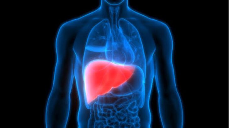
The liver stands as the largest internal solid organ in the human body, serving as a master metabolic processing center that regulates nutrient storage, filters toxins from the blood, synthesizes vital plasma proteins, and produces bile to aid digestion. Under healthy physiological conditions, hepatocytes—the primary functional cells of the liver—maintain a tightly regulated balance of lipid metabolism, processing dietary fats and transporting triglycerides efficiently without accumulating excess fat within their cytoplasm. However, when systemic metabolic imbalances, insulin resistance, or chronic toxins overwhelm this delicate lipid homeostasis, fat droplets begin to accumulate abnormally inside the hepatocytes. This pathological fat infiltration is the defining hallmark of fatty liver disease, a remarkably common and potentially progressive condition that ranges from benign fat accumulation to severe liver destruction. Understanding the underlying mechanisms and natural history of this condition reveals how a metabolic disturbance can slowly transform a healthy organ into a site of severe fibrosis.
Primary Classifications: NAFLD, MASH, and Alcoholic Liver Disease
Fatty liver disease is broadly categorized into two major clinical classifications based on its primary etiology, though both share remarkably similar histological pathways of progressive liver injury.
-
Metabolic Dysfunction-Associated Steatotic Liver Disease: The most prevalent form is metabolic dysfunction-associated steatotic liver disease, closely linked to the global rise in obesity, type 2 diabetes mellitus, dyslipidemia, and metabolic syndrome. Within this category, simple steatosis—where fat accumulates without significant inflammation—can progress into metabolic dysfunction-associated steatohepatitis, historically known as NASH.
-
Alcohol-Related Liver Disease: The second major category is alcohol-related liver disease, which is directly precipitated by chronic, excessive ethanol consumption that disrupts hepatic lipid metabolism, generates oxidative stress, and triggers severe cellular inflammation. Both pathways can drive progressive structural damage if the underlying triggers remain unaddressed.
The Spectrum of Progression: From Steatosis to Steatohepatitis
To understand how fatty liver disease advances, one must examine the histological stages that define its clinical progression over years or decades.
-
Simple Steatosis: The initial stage is simple hepatic steatosis, defined by the accumulation of fat in more than five percent of hepatocytes. While historically viewed as a benign, non-progressive condition, simple steatosis creates a vulnerable metabolic environment within the liver.
-
Steatohepatitis and Oxidative Stress: Over time, secondary hits—including lipotoxicity, insulin resistance, oxidative stress, and the release of pro-inflammatory cytokines like tumor necrosis factor-alpha—trigger the transition to steatohepatitis. In this inflamed state, hepatocytes experience ballooning degeneration, cellular injury, and localized immune cell infiltration as white blood cells flood the liver tissue to combat cellular debris, initiating the scarring process.
Fibrosis Development and the Path to Cirrhosis
As chronic inflammation persists within the hepatic parenchyma, the ongoing cellular damage activates specialized cells known as hepatic stellate cells residing in the space of Disse. Under normal conditions, these cells store vitamin A and remain quiescent; however, cytokines released during chronic steatohepatitis stimulate them to transform into active myofibroblasts. These activated myofibroblasts begin churning out massive quantities of extracellular matrix proteins, including collagen scar tissue, laying down fibrous bands that weave through the liver architecture.
-
Bridging Fibrosis: As this fibrotic scarring intensifies over years, it forms bridging connections between portal tracts and central veins, distorting the normal microvascular architecture.
-
Cirrhosis: Eventually, widespread scar tissue replaces functional hepatic parenchyma, creating regenerative nodules and culminating in cirrhosis. At this advanced stage, the liver becomes hard, shrunken, and structurally compromised, severely impairing blood flow through the portal vein system and precipitating portal hypertension.
Clinical Presentation and Silent Disease Progression
One of the most challenging aspects of fatty liver disease is its insidious, silent nature during the early and intermediate stages of progression. Because the liver possesses an extraordinary functional reserve and lacks pain receptors within its parenchyma—relying for pain sensation only on its outer fibrous capsule—individuals with simple steatosis or early steatohepatitis frequently remain completely asymptomatic for decades. When symptoms do eventually arise as fibrosis advances, they are often vague and non-specific, including persistent fatigue, a dull, aching discomfort in the right upper quadrant of the abdomen, and generalized malaise. Consequently, many patients are diagnosed incidentally during routine blood work revealing elevated liver enzymes or through abdominal imaging performed for unrelated medical reasons, underscoring the importance of proactive screening in high-risk metabolic populations.
Diagnostic Investigations: Biomarkers, Imaging, and Biopsy
Detecting and staging fatty liver disease requires a combination of non-invasive laboratory tests, advanced imaging modalities, and occasionally histological tissue evaluation.
-
Laboratory Screening: Blood panels evaluate liver enzymes—such as alanine aminotransferase and aspartate aminotransferase—alongside metabolic profiles, though normal enzyme levels do not definitively rule out advanced fibrosis.
-
Imaging Modalities: Diagnostic imaging plays a pivotal role; ultrasound can readily identify moderate to severe steatosis, while specialized techniques like vibration-controlled transient elastography measure liver stiffness and fat content quantitatively, providing a non-invasive assessment of fibrosis severity.
-
Histological Confirmation: When non-invasive results are ambiguous or clinical trials require definitive staging, a percutaneous liver biopsy remains the gold standard, allowing pathologists to examine tissue under a microscope to grade inflammation and stage the exact extent of scar tissue accumulation.
Management Strategies and Lifestyle Interventions
Because there is currently no single pharmacological cure for fatty liver disease, management focuses primarily on halting or reversing disease progression through targeted lifestyle modifications and addressing root metabolic risk factors.
-
Weight Loss and Dietary Modification: For metabolic dysfunction-associated steatotic liver disease, achieving sustained weight loss through a balanced, calorie-restricted diet—such as the Mediterranean diet rich in antioxidants, healthy fats, and low in refined sugars and fructose—is the cornerstone of therapy. Gradual weight loss of seven to ten percent of total body weight has been proven clinically to reduce hepatic steatosis, decrease inflammation, and even reverse early fibrosis.
-
Exercise and Pharmacotherapy: Regular aerobic and resistance exercise improves systemic insulin sensitivity independently of weight loss. Furthermore, emerging pharmacotherapies targeting metabolic pathways, GLP-1 receptor agonists, and specialized antioxidants are increasingly utilized by hepatologists to treat high-risk patients with progressive steatohepatitis and advanced fibrosis, preventing the ultimate transition to liver failure.
Conclusion
Fatty liver disease is a progressive metabolic condition characterized by abnormal lipid accumulation within hepatocytes, carrying the potential to advance from simple steatosis to severe steatohepatitis, extensive fibrosis, and life-threatening cirrhosis. By examining how systemic insulin resistance, oxidative stress, and chronic inflammation drive hepatic stellate cell activation and scar tissue formation, the critical importance of early detection becomes undeniable. Through proactive screening in metabolic populations, non-invasive elastography diagnostics, targeted lifestyle interventions focusing on weight loss and nutrition, and modern pharmacological management, healthcare providers can successfully halt disease progression, promote tissue healing, and preserve long-term hepatic health.