Understanding Heart Failure With Preserved Ejection Fraction
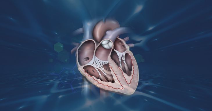
The human cardiovascular apparatus maintains a continuous, tightly regulated cycle of contraction and relaxation to secure adequate perfusion throughout all systemic tissues. Clinical practitioners traditionally judged the efficacy of this cardiac cycle primarily via the calculation of ejection fraction, a metric denoting the proportion of blood expelled from the left ventricular chamber during the contraction phase known as systole. For generations, cardiac failure was tightly linked to a severely reduced ejection fraction, denoting a flaccid, dilated pump incapable of driving sufficient volume forward. Nevertheless, contemporary cardiology recognizes a distinct and equally perilous clinical entity designated as heart failure with preserved ejection fraction, formerly termed diastolic failure. In this paradoxical condition, the muscular walls contract with robust force, maintaining an ejection fraction above fifty percent, yet fail to relax and accommodate incoming blood properly during the resting phase termed diastole. This profound inability to accept returning venous blood initiates a destructive hemodynamic cascade defining the disease.
The Pathophysiology of Diastolic Dysfunction and Ventricular Stiffness
To fully grasp the mechanical failures underpinning this disorder, one must evaluate the intricate biological controls governing myocardial relaxation and chamber compliance. Diastole functions as an active, energy-demanding process termed lusitropy, requiring high quantities of adenosine triphosphate to uncouple actin and myosin filaments following cellular contraction. In this condition, this active relaxation step is severely compromised. Furthermore, the passive elasticity of the ventricular wall undergoes pathological remodeling. Chronic hemodynamic stress stimulates resident fibroblasts to synthesize excessive collagen, generating dense extracellular matrix fibrosis that physically stiffens the muscular tissue. Concurrently, molecular changes occur within titin, a giant spring-like protein regulating cellular elasticity. Pathological structural modifications render these titin filaments abnormally rigid, yielding a thickened, hypertrophied, and unyielding ventricle requiring dangerously high pressures simply to fill with an appropriate volume of blood.
Systemic Inflammation and Microvascular Endothelial Dysfunction
Modern cardiovascular investigation has shifted the paradigm regarding this illness, demonstrating that it is not merely an isolated myopathic defect, but rather a complex systemic syndrome. The condition is driven heavily by the convergence of metabolic comorbidities, specifically severe adiposity, chronic hypertension, and type 2 diabetes mellitus. These concurrent metabolic disturbances provoke a persistent, low-grade inflammatory state throughout the organism. This circulating inflammatory milieu inflicts severe injury upon the delicate endothelial cells lining the coronary microvasculature. When these microscopic vessels become dysfunctional, they lose their capacity to synthesize adequate nitric oxide, a messenger vital for maintaining vascular tone and myocardial relaxation. The resultant depletion of nitric oxide pathways suppresses protein kinase G activity inside cardiac cells, directly promoting further cellular hypertrophy, exacerbating titin stiffness, and locking the heart into a progressive structural decline.
Clinical Presentation and Hemodynamic Consequences
The functional fallout of this stiffened, non-compliant chamber manifests through a specific constellation of disabling symptoms limiting daily activities. Because the left ventricle cannot expand to receive oxygenated pulmonary blood without elevated filling pressures, this immense retrograde pressure backs up directly into the left atrium and pulmonary venous bed. This pulmonary congestion provokes the hallmark symptom of exertional dyspnea, wherein patients suffer severe shortness of breath during mild physical tasks. As the illness progresses, individuals frequently develop orthopnea, needing multiple pillows for nighttime comfort, alongside paroxysmal nocturnal dyspnea that jolts them awake gasping for air. Moreover, because the rigid heart cannot adequately increase its stroke volume during physical exertion, sufferers endure profound exercise intolerance and overwhelming fatigue, frequently accompanied by chronotropic incompetence, an inability to elevate heart rate sufficiently during activity.
Diagnostic Evaluation and Echocardiographic Imaging
Diagnosing this intricate pathology requires a meticulous multimodal assessment, because the standard metric of a normal ejection fraction can mask severe underlying damage. Clinicians rely heavily on advanced echocardiography to identify subtle structural and functional irregularities. While overall contractile performance appears visually intact, Doppler interrogation unveils crucial markers of diastolic dysfunction, particularly an elevated ratio of early mitral inflow velocity to mitral annular early diastolic velocity, a parameter correlating tightly with elevated left ventricular filling pressures. Ultrasounds also frequently reveal an enlarged left atrium, acting as a biological barometer reflecting chronic pressure overload over time. To support findings, physicians assess circulating biomarkers like B-type natriuretic peptides, secreted by stretched cardiac tissues, though values can appear deceptively normal in severely obese individuals, complicating detection.
Advanced Hemodynamic Assessments and Right Heart Catheterization
When non-invasive imaging and biomarker profiles remain inconclusive, cardiovascular specialists must employ invasive hemodynamic testing to secure a definitive diagnosis. The absolute gold standard for confirming this syndrome involves executing a right heart catheterization, where a specialized balloon catheter travels through the venous system into the right heart chambers and out into the pulmonary vasculature. This procedure enables clinicians to precisely measure pulmonary capillary wedge pressure, mirroring pressures trapped within the left atrium and ventricle. Because many patients display entirely normal hemodynamics while resting quietly on an examination table, clinicians frequently perform this test dynamically while the patient exercises on a recumbent bicycle within the catheterization suite. Observing a pathological spike in filling pressures during mild exertion offers definitive confirmation of the stiffened ventricular phenotype.
Therapeutic Interventions and Pharmacological Paradigm Shifts
Historically, the pharmacological landscape for this condition remained bleak, featuring decades of clinical trials where traditional heart failure agents failed to improve survival, leaving physicians reliant on diuretics to manage fluid overload. However, a major breakthrough revolutionized management with the introduction of sodium-glucose cotransporter-2 inhibitors, such as empagliflozin and dapagliflozin. Initially engineered as oral anti-diabetic treatments, these drugs demonstrated an unprecedented ability to reduce cardiovascular hospitalizations and mortality in this specific cohort. While exact cardioprotective mechanisms are explored, they foster favorable osmotic diuresis, optimize cellular energy metabolism, and directly mitigate systemic inflammation and fibrosis. Alongside these novel drugs, clinicians utilize mineralocorticoid receptor antagonists like spironolactone to counter fibrotic remodeling, while actively treating hypertension and atrial fibrillation to optimize filling intervals.
Conclusion
Heart failure with preserved ejection fraction constitutes one of the most challenging epidemics in contemporary cardiovascular practice, fueled by an aging population and rising metabolic disease rates. Recognizing that this disorder extends beyond a simple mechanical filling deficit to involve systemic inflammation, microvascular dysfunction, and complex cellular remodeling clarifies the true severity of the condition. Through the integration of advanced echocardiography, dynamic invasive testing, and the application of targeted pharmacological therapies addressing both the myocardium and systemic inflammation, healthcare practitioners can alter disease trajectories and restore functional capacity.