What Is Endometriosis and How Can It Affect the Reproductive System?
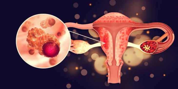
Under normal physiological conditions, the structure and thickness of the endometrium fluctuate continuously throughout the menstrual cycle, governed by cyclic variations in pituitary gonadotropins (follicle-stimulating hormone and luteinizing hormone) and ovarian steroid hormones (estrogen and progesterone). During the proliferative phase, rising circulating estradiol concentrations stimulate rapid cellular mitosis, glandular elongation, and vascular growth within the functional layer of the endometrium. Following ovulation, progesterone secreted by the corpus luteum converts the tissue into a receptive, secretory environment suitable for embryo implantation. If fertilization does not take place, the withdrawal of progesterone triggers enzymatic degradation, focal tissue necrosis, and shedding of the functionalis layer during menstruation. This ectopic tissue responds to cyclic hormonal signaling, driving persistent localized inflammation, pelvic pain, extensive fibrosis, and significant reproductive dysfunction.
Etiological Hypotheses and Cellular Pathogenesis
The precise molecular and cellular mechanisms responsible for the initiation and establishment of ectopic endometrial implants remain a subject of extensive investigation, with multiple interlinked theories explaining its pathogenesis. The most widely accepted foundation is Sampson’s Theory of Retrograde Menstruation, which posits that during menses, endometrial tissue fragments flow backward through the Fallopian tubes into the peritoneal cavity. However, because retrograde flux occurs in up to ninety percent of menstruating women, additional cellular defects are necessary for ectopic implantation to take place. These systemic and local factors include altered cell adhesion dynamics, defective peritoneal immune clearance, heightened local angiogenesis, and somatic genetic mutations. Alternative mechanisms include Coelomic Metaplasia—the transformation of primitive peritoneal mesothelial cells into endometrial-like epithelium under hormonal or inflammatory stimuli—and Vascular/Lymphatic Dissemination, which accounts for rare extra-pelvic endometriotic deposits observed in distant organs such as the lungs, brain, or surgical scars.
Inflammatory Signaling, Immune Dysfunction, and Lesion Evolution
Once ectopic endometrial cells attach to pelvic peritoneal surfaces or ovarian tissues, they establish a self-sustaining pro-inflammatory microenvironment. Natural killer (NK) cell cytotoxicity and peritoneal macrophage phagocytic activity are diminished in affected individuals, allowing viable endometrial cells to escape immune degradation. Ectopic tissues express abnormal intracellular levels of aromatase and 17-beta-hydroxysteroid dehydrogenase type 1, driving local biosynthesis of estradiol directly within the lesions. Furthermore, local estrogen production upregulates Cyclooxygenase-2 (COX-2) expression, escalating the release of Prostaglandin E2 (PGE2). This creates a positive feedback loop: PGE2 further amplifies aromatase activity, enhancing local estrogen concentrations while recruiting mast cells, macrophages, and pro-inflammatory cytokines such as Interleukin-1 beta (IL-1b), Interleukin-6 (IL-6), Interleukin-8 (IL-8), and Tumor Necrosis Factor-alpha (TNF-a). This chronic inflammatory milieu drives neo-angiogenesis via Vascular Endothelial Growth Factor (VEGF) and stimulates nerve fiber growth (neurogenesis), transforming ectopic implants into vascularized, pain-generating nodular lesions.
Impact on Female Reproductive Architecture and Ovarian Function
Endometriosis exerts severe, multi-faceted structural and functional disruptions across the female reproductive system. Pelvic lesions undergo cyclic shedding, bleeding, and incomplete clearing, resulting in repeated localized micro-hemorrhages. Over time, the chemical irritation caused by free blood within the peritoneal cavity initiates severe inflammatory cascades that promote excessive deposition of fibrinous exudate, leading to dense fibrous adhesions. These mechanical adhesions lock the uterus, Fallopian tubes, ovaries, and pouch of Douglas into distorted anatomical configurations, obliterating normal spatial relationships and physically obstructing tubal motility and ovum pickup. Within the ovaries, deep invagination of ectopic tissue generates ovarian Endometriomas (commonly known as "chocolate cysts"). These chronic cysts contain dark, oxidized blood products that exert mechanical pressure and induce localized oxidative stress on surrounding normal tissue, progressively diminishing the ovarian reserve by accelerating primordial follicle depletion and compromising oocyte quality.
Mechanisms of Infertility and Gamete Quality Degradation
Infertility is one of the most severe clinical sequelae of endometriosis, affecting up to thirty to fifty percent of women diagnosed with the condition. While anatomical distortion from pelvic adhesions and tubal blockage provides a clear mechanical cause for subfertility, endometriosis also impairs fertility in early-stage disease without mechanical obstruction. The elevated concentration of reactive oxygen species (ROS), inflammatory cytokines, and proteolytic enzymes within the peritoneal fluid damages both sperm and oocyte integrity. Spermatozoa traversing the uterine and peritoneal microenvironments experience impaired motility, reduced acrosome reaction capacity, and increased DNA fragmentation. Simultaneously, chronic intra-follicular inflammation alters granulosa cell function, leading to impaired steroidogenesis, poor oocyte maturation, compromised zona pellucida penetration, and defective embryonic cleavage. Furthermore, progesterone resistance within the eutopic endometrial lining reduces expression of crucial implantation markers (such as alpha-v-beta-3 integrins and HOXA10), impairing endometrial receptivity and preventing successful blastocyst implantation.
Clinical Manifestations, Neurogenic Pain, and Diagnostic Protocols
Patients frequently experience severe Dysmenorrhea (painful menstruation), Deep Dyspareunia (pain during sexual intercourse), Dyschezia (painful bowel movements during menses), Dysuria (painful urination), and non-menstrual Chronic Pelvic Pain. Pain mechanics involve both nociceptive activation from inflammatory mediators and neurogenic mechanisms resulting from direct infiltration of sensory nerve fibers within deep infiltrating endometriosis (DIE) nodules, leading to central sensitization over time. Diagnosing endometriosis requires combining comprehensive clinical history, pelvic examination, and advanced imaging modalities. Transvaginal Ultrasonography (TVUS) with deep-endometriosis protocols and Magnetic Resonance Imaging (MRI) exhibit high sensitivity for detecting ovarian endometriomas and deep pelvic infiltrating nodules. However, Diagnostic Laparoscopy paired with histological confirmation remains the definitive diagnostic gold standard for visualizing superficial peritoneal implants and staging disease severity using the Revised American Society for Reproductive Medicine (rASRM) classification system.
Therapeutic Management Strategies and Clinical Interventions
Managing endometriosis requires an individualized, multidisciplinary approach focused on suppressing symptoms, preserving or restoring fertility, and halting disease progression. Medical therapies primarily utilize hormonal suppression to create a hypo-estrogenic or hyper-progestogenic environment that induces atrophy of both ectopic and eutopic endometrial tissue. First-line agents include Combined Oral Contraceptives and continuous Progestins (such as dienogest or medroxyprogesterone acetate). Second-line therapies utilize Gonadotropin-Releasing Hormone (GnRH) agonists or oral GnRH antagonists, which downregulate the hypothalamic-pituitary-ovarian axistoinduce temporary therapeutic menopause; these are routinely paired with "add-back" low-dose estrogen/progestin therapy to prevent bone mineral density lossand vasomotor symptoms. For patients with severe pain refractory to medication, mechanical tubal occlusion, or large endometriomas, Surgical Intervention via laparoscopic excision (peritoneal lesion resection and endometrioma cystectomy) restores pelvic anatomy and alleviates pain. In infertile individuals with advanced structural disease or low ovarian reserve, Assisted Reproductive Technologies (ART), specifically In Vitro Fertilization (IVF), provide the most effective path to achieving pregnancy.
Conclusion
Endometriosis is a complex, estrogen-dependent inflammatory disorder characterized by ectopic endometrial tissue proliferation, chronic pelvic inflammation, and severe structural remodeling. Understanding how retrograde transport, local estrogen biosynthesis, and immune clearance defects interact clarifies the progression from superficial peritoneal implants to deep infiltrating nodules, dense pelvic adhesions, and ovarian endometriomas. Utilizing high-resolution transvaginal ultrasound, pelvic MRI, and diagnostic laparoscopy enables clinical teams to map lesion distribution and grade disease severity accurately. Through targeted hormonal suppression, laparoscopic excision, and advanced assisted reproductive technologies, healthcare professionals can effectively suppress neuroinflammatory pain, restore pelvic structural integrity, and optimize long-term reproductive outcomes for affected individuals.