What Is Pulmonary Valve Stenosis and How Does It Affect Blood Flow?
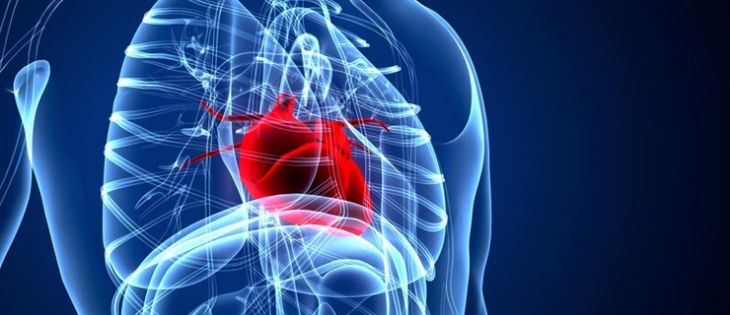
In normal human cardiovascular physiology, four distinct heart valves regulate unidirectional blood flow through sequential cardiac chambers and systemic vessels. The pulmonary valve is a tri-leaflet semilunar structure positioned at the junction between the right ventricular outflow tract (RVOT) and the main pulmonary artery. During ventricular systole, increased pressure inside the right ventricle forces the three thin, pliable pulmonary valve cusps to separate smoothly toward the arterial wall, allowing deoxygenated blood to flow into the pulmonary circulation for gas exchange. During diastole, as right ventricular pressure drops below pulmonary arterial pressure, the valve cusps coapt cleanly in the center of the vessel trunk to prevent retrograde regurgitation into the ventricle. Pulmonary Valve Stenosis (PVS) is a structural cardiovascular lesion defined by restricted opening or anatomical narrowing of the pulmonary valve aperture. This mechanical obstruction impedes systolic blood ejection, generating a substantial pressure gradient between the right ventricle and the main pulmonary artery, which alters structural wall geometry and alters right heart hemodynamics.
Etiological Classifications and Structural Pathology
Pulmonary valve stenosis arises primarily as a congenital heart anomaly, representing roughly eight to ten percent of all congenital cardiac defects. Anatomically, outflow tract obstructions are classified into three structural sub-types based on the exact site of narrowing: valvular, subvalvular (infundibular), and supravalvular. Typical valvular pulmonary stenosis results from incomplete separation or commissural fusion of the valve cusps during embryonic cardiac development, creating a dome-like valve structure with a small, unyielding central aperture. A distinct histological variant is the dysplastic pulmonary valve, which features thickened, rigid, nodular leaflets composed of excessive fibrous and myxoid matrix tissue without significant commissural fusion. Dysplastic valves are strongly linked to inherited genetic conditions, most notably Noonan syndrome. Less commonly, acquired forms of pulmonary valve narrowing develop secondary to carcinoid heart disease—in which circulating neuroendocrine peptides cause fibrous plaque deposition on endocardial surfaces—or late structural sequelae of severe rheumatic heart disease.
Pathophysiology of Pressure Overload and Ventricular Hypertrophy
The primary mechanical anomaly in pulmonary valve stenosis is fixed resistance to systolic blood ejection from the right ventricle. To force blood across the narrowed valvular orifice and maintain sufficient pulmonary blood flow, the right ventricle must generate elevated peak systolic pressures. This chronic intraventricular elevation produces a persistent pressure gradient across the pulmonary valve during ejection, which directly reflects the clinical severity of the stenosis. Applying Laplace's law of wall stress—where circumferential tension relates directly to intraventricular pressure and radius while inversely relating to wall thickness—the right ventricle adapts to chronic pressure overload through concentric myocardial hypertrophy. Cardiomyocytes synthesize new sarcomeres in parallel, thickening the muscular right ventricular wall and increasing myocardial mass to reduce wall stress back toward baseline levels.
Hemodynamic Progression to Right Ventricular Failure
Although concentric hypertrophy temporarily compensates for elevated outflow resistance and preserves resting stroke volume, uncorrected pressure overload eventually triggers secondary hemodynamic decompensation. As right ventricular wall thickness increases, myocardial compliance drops, creating diastolic dysfunction. The non-compliant ventricular wall requires higher filling pressures during diastole, causing right atrial enlargement, elevated right atrial contraction pressures, and systemic venous hypertension. Concurrently, hyper-thickened myocardium exhibits increased metabolic demand, making subendocardial tissue susceptible to chronic ischemia due to microvascular compression during high-tension systole. Over time, persistent hypoxic stress and myocardial fibrosis exhaust contractile reserves, precipitating systolic heart failure. As ejection fraction declines, end-systolic volume rises, causing right ventricular dilation, secondary tricuspid ring dilation with functional regurgitation, systemic venous stasis, and peripheral organ congestion.
Clinical Presentation, Auscultatory Findings, and Symptomatology
The clinical presentation of pulmonary valve stenosis varies depending on the degree of mechanical obstruction and the patient's age. Mild pulmonary stenosis (peak transvalvular Doppler gradient under thirty millimeters of mercury) usually causes no noticeable symptoms and is often discovered incidentally during routine pediatric or clinical check-ups. Moderate-to-severe stenosis (peak gradients exceeding fifty to eighty millimeters of mercury) produces symptoms caused by fixed cardiac output and systemic venous backpressure. Affected individuals report exertional dyspnea, fatigue, impaired exercise capacity, and ischemic chest discomfort during physical effort. Severe decompensated cases display overt systemic venous congestion, including peripheral edema, hepatomegaly, ascites, and prominent "a" waves on jugular venous pulse examination. Auscultation characteristically reveals a harsh crescendo-decrescendo systolic ejection murmur at the upper left sternal border in the second intercostal space, often accompanied by an early ejection click in flexible, pliable valvular stenosis that decreases in intensity during inspiration.
Diagnostic Evaluation Protocols and Multimodal Imaging
Accurate diagnosis and functional grading of pulmonary valve stenosis rely on non-invasive cardiovascular imaging techniques. Transthoracic Echocardiography (TTE) integrated with spectral continuous-wave Doppler serves as the primary diagnostic tool. Continuous-wave Doppler records peak flow velocities across the stenotic valve, enabling clinicians to calculate peak and mean pressure gradients using the simplified Bernoulli equation. Echocardiography also assesses valve cusp thickness, commissural fusion, right ventricular wall thickness, chamber dimensions, right atrial enlargement, and concomitant tricuspid valve function. Cardiac Magnetic Resonance (CMR) imaging acts as an essential secondary imaging technique, providing precise volumetric measurements, right ventricular ejection fraction calculations, and tissue characterization without technical acoustic window limitations. Diagnostic cardiac catheterization provides direct invasive pressure tracings within the right ventricle and pulmonary artery when non-invasive measurements are inconclusive or when planning therapeutic catheter interventions.
Therapeutic Interventions and Surgical Management Strategies
Therapeutic management of pulmonary valve stenosis is guided by the magnitude of the transvalvular pressure gradient, valve morphology, and clinical symptoms. Mild, asymptomatic valvular stenosis requires no invasive treatment and is managed conservatively with routine clinical follow-up, as gradient progression is uncommon in older children and adults. For patients with severe valvular stenosis—defined as a peak Doppler gradient over fifty millimeters of mercury with symptoms, or an asymptomatic peak gradient exceeding sixty to eighty millimeters of mercury—Percutaneous Balloon Pulmonary Valvuloplasty(BPV) is the gold standard therapeutic procedure. During BPV, a balloon-tipped catheter is navigated across the stenotic valve orifice and inflated to tear fused commissures, significantly enlarging the valve area and relieving outflow obstruction with minimal risk. In patients with dysplastic valve cusps, marked subvalvular infundibular stenosis, or failed percutaneous balloon dilation, open surgical repair—including pulmonary valvotomy, infundibular muscle resection, or transannular patch enlargement—is indicated to restore unhindered right ventricular ejection.
Conclusion
Pulmonary Valve Stenosis is an important structural cardiovascular disorder defined by right ventricular outflow obstruction, leading to chronic pressure overload and adaptive concentric right ventricular hypertrophy. Analyzing how valvular narrowing elevates intraventricular pressures explains the pathophysiological transition from early compensated hypertrophy to diastolic stiffness, right ventricular enlargement, and systemic venous congestion. Combining continuous-wave Doppler echocardiography, cardiac magnetic resonance, and catheterization tracings enables clinical teams to evaluate gradient severity and select optimal management pathways. By utilizing timely percutaneous balloon pulmonary valvuloplasty or targeted surgical repair, clinical teams can effectively relieve outflow obstruction, normalize intraventricular pressures, and safeguard long-term cardiovascular performance for affected individuals.