How Acoustic Imaging Technology Could Help Researchers Examine Tissues Without Surgery
Examining the body's internal tissues is an important part of medical research and disease diagnosis. Doctors and researchers need to understand how organs, muscles, blood vessels, and other structures look and function. Traditionally, some detailed tissue examinations have required surgical procedures or the collection of tissue samples. However, advances in imaging technology are creating new ways to study the body without making surgical cuts.
Acoustic imaging is one area receiving attention. It uses sound waves to produce images of internal structures and may help researchers examine tissues without surgery. Modern techniques are also exploring how sound signals can reveal information about tissue stiffness, movement, and other physical properties.
What Is Acoustic Imaging Technology?
Acoustic imaging is a method of creating images using sound waves. In medical applications, it commonly involves ultrasound technology, which sends high-frequency sound waves into the body.
When these waves reach different tissues, some of the sound energy is reflected back. A sensor called a transducer detects the returning signals. A computer then processes them to create images of structures inside the body.
Different tissues reflect sound waves in different ways. These differences allow imaging systems to distinguish organs, fluids, blood vessels, and other structures.
Unlike X-rays and CT scans, ultrasound does not use ionizing radiation. This makes it useful for several types of medical examinations.
How Does Acoustic Imaging Examine Body Tissues?
Acoustic imaging systems use several steps to examine internal structures.
Sending Sound Waves Into the Body
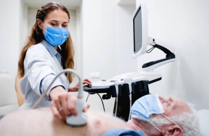
A healthcare professional places an ultrasound transducer against the skin, usually with a special gel that helps sound waves travel between the device and the body.
The transducer sends high-frequency sound waves into the tissues.
Receiving Reflected Signals
As sound waves travel through the body, they encounter structures with different physical properties. Some waves are reflected toward the transducer.
The device records these returning signals and measures how they change.
Creating Internal Images
A computer processes the signals to produce images of the examined area. These images can show the size, shape, position, and movement of different structures.
Studying Tissue Characteristics
Advanced acoustic imaging techniques may provide information beyond basic tissue appearance. Researchers are studying methods that can estimate tissue stiffness, detect movement, and examine changes in blood flow.
These measurements may help researchers understand how certain diseases affect tissues.
Different Types of Acoustic Imaging Techniques
Several imaging methods use sound waves to examine the body. Each technique provides different information.
Conventional Ultrasound Imaging
Conventional ultrasound creates real-time images of internal organs and tissues. It is widely used to examine the abdomen, thyroid, heart, blood vessels, and developing babies during pregnancy.
It can help identify changes such as swelling, fluid-filled areas, cysts, and unusual tissue structures.
Elastography
Elastography is an advanced ultrasound technique that estimates tissue stiffness or elasticity.
Healthy and diseased tissues can have different mechanical properties. For example, some forms of liver disease cause scar tissue to develop, making the liver stiffer.
Researchers and healthcare professionals use elastography to assess tissue changes without always needing a biopsy.
Doppler Ultrasound
Doppler ultrasound examines the movement of blood through blood vessels. It measures changes in sound-wave frequency caused by moving blood cells.
This technique helps assess blood flow and can identify certain circulation problems.
Photoacoustic Imaging
Photoacoustic imaging combines light and sound. Short pulses of laser light are absorbed by tissues, causing small temperature changes and producing sound waves.
Special sensors detect these waves and use them to create images. Researchers are studying this method to examine blood vessels, oxygen levels, and tissue characteristics.
However, many photoacoustic applications remain under investigation and are not yet routine clinical tests.
How Could Acoustic Imaging Help Researchers Study Diseases?
Researchers are exploring how advanced sound-based imaging could improve the understanding of different diseases.
Studying Liver Conditions
Liver diseases can cause inflammation, fat accumulation, and fibrosis. Elastography can estimate liver stiffness and help assess certain changes associated with fibrosis.
Researchers are also investigating ways to combine acoustic measurements with other imaging information to improve disease assessment.
Examining Tumors
Tumors may differ from surrounding tissues in their stiffness, structure, and blood supply. Acoustic imaging research aims to identify these differences and understand how tumors change over time.
Some techniques may also help researchers examine how tissues respond to cancer treatments.
However, acoustic imaging findings alone cannot confirm that a tissue abnormality is cancerous.
Understanding Heart and Blood Vessel Conditions
Ultrasound is already important in cardiovascular examinations. It helps doctors observe heart movement, valve function, and blood flow.
Advanced research is examining how improved acoustic techniques could provide more detailed information about blood vessels and changes in heart tissue.
Studying Muscle and Joint Disorders
Sound-based imaging can examine muscles, tendons, ligaments, and other soft tissues. Researchers use ultrasound to study tissue movement and changes associated with injuries or certain muscle disorders.
This can provide useful information without requiring surgery.
What Are the Benefits of Examining Tissues Without Surgery?
Non-invasive imaging can offer several advantages for medical research and patient care.
No surgical cuts: Many ultrasound examinations can be performed without making an incision.
No ionizing radiation: Ultrasound uses sound waves rather than X-rays.
Real-time information: Researchers can observe movement and certain changes as they happen.
Repeated examinations: Some techniques can be used repeatedly to monitor changes over time.
Reduced discomfort: Most external ultrasound examinations are painless or cause only mild pressure.
Potentially lower costs: Ultrasound equipment and examinations may be less expensive than some advanced imaging methods, depending on the setting.
These advantages make acoustic imaging valuable for many research and clinical applications.
What Are the Limitations of Acoustic Imaging?
Acoustic imaging also has limitations. Sound waves do not travel equally well through every type of body tissue. Bones and air can block or weaken ultrasound signals, making some areas difficult to examine.
Image quality can also depend on the equipment, operator experience, and the patient's body characteristics.
In addition, acoustic imaging cannot provide all the information that a tissue biopsy can offer. A biopsy allows specialists to examine cells directly and identify certain cellular or molecular changes.
For this reason, doctors may combine ultrasound with MRI, CT scans, laboratory tests, or biopsy when more detailed information is needed.
What Does the Future of Acoustic Imaging Look Like?
Researchers are working to improve sound-based imaging through artificial intelligence, advanced sensors, and more powerful image-processing methods.
AI systems may help analyze complex ultrasound images, identify unusual tissue patterns, and reduce some differences in image interpretation. New transducer designs may also improve image quality and make certain examinations easier.
Another research direction involves combining acoustic imaging with other technologies to obtain more information from the same examination.
These developments could make non-invasive tissue assessment more useful in the future. However, new methods must undergo proper clinical testing before they can be considered reliable for routine diagnosis.
FAQs
Is acoustic imaging the same as ultrasound?
Ultrasound is one of the most widely used forms of medical acoustic imaging. The broader field also includes advanced techniques such as elastography and photoacoustic imaging.
Can acoustic imaging examine tissues without surgery?
Yes. Many acoustic imaging methods examine internal tissues through the skin without surgical cuts. However, some conditions may still require a biopsy or another medical test.
Can acoustic imaging detect cancer?
Acoustic imaging can identify unusual tissue structures and help doctors examine suspicious areas. However, it cannot confirm every cancer diagnosis. Additional testing, including a biopsy, may be necessary.
Is acoustic imaging safe?
Medical ultrasound does not use ionizing radiation and is generally considered safe when performed appropriately by trained professionals.
Conclusion
Acoustic imaging technology is changing how researchers and healthcare professionals examine internal tissues. By using sound waves, these methods can produce images and provide information about tissue structure, stiffness, movement, and blood flow without requiring surgery in many cases.
Conventional ultrasound is already widely used, while advanced methods such as elastography and photoacoustic imaging continue to expand research possibilities. Although these technologies cannot replace every medical test or biopsy, ongoing developments may make non-invasive tissue examination more detailed and useful in the future.