What Is Endocarditis and How Can It Affect the Heart?
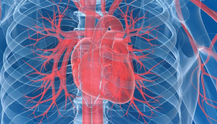
The human heart is protected by three distinct tissue layers, the innermost of which is the endocardium—a smooth, glistening membrane of endothelial cells that lines the internal cavities of the heart chambers and covers the delicate leaflets of all four cardiac valves. Under normal physiological circumstances, this continuous endothelial lining provides a slick, non-thrombogenic surface that allows blood to flow smoothly without adhering to structural walls. However, when microscopic disruptions occur on this delicate surface, or when turbulent blood flow damages the endothelial cells, microscopic sites of sterile platelet and fibrin deposition can form. If circulating bacteria or fungi enter the bloodstream and encounter these vulnerable patches, they can attach, colonize, and multiply, precipitating a dangerous and potentially life-threatening infection known as infective endocarditis. Understanding the complex pathogenesis of this condition reveals how a systemic microbial invasion can severely compromise structural cardiac integrity.
Pathogenesis: Endothelial Disruption and Vegetative Formation
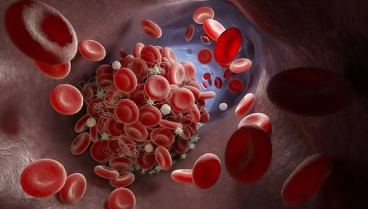
To understand how infective endocarditis develops, one must examine the sequential pathological events that transform a minor bloodstream exposure into a destructive cardiac infection. The initial prerequisite is non-bacterial thrombotic endocarditis, where mechanical stress, congenital valve abnormalities, or degenerative wear and tear roughen the endothelial lining of a heart valve, causing tiny clots composed of platelets and fibrin to accumulate. During episodes of transient bacteremia—which can occur during routine dental procedures, invasive medical interventions, or even daily activities like tooth brushing—circulating microorganisms passing through the heart collide with these sterile micro-clots. The invading bacteria adhere to the exposed matrix proteins, engulf themselves in a protective fibrin and platelet meshwork, and begin multiplying rapidly, creating characteristic masses known as vegetations. These fragile, microbe-laden growths adhere tenaciously to the valve leaflets, disrupting normal closure and serving as protected microbial reservoirs.
Hemodynamic Consequences and Valvular Destruction
The presence of growing vegetations on the heart valves inflicts profound mechanical and structural damage upon the cardiovascular system. As the microbial masses expand, they physically interfere with the normal coaptation of the valve leaflets, preventing them from sealing tightly and resulting in acute or progressive valvular regurgitation. Furthermore, the relentless bacterial proliferation and the host's localized inflammatory response cause enzymatic destruction of the delicate valve tissue, leading to leaflet perforation, chordae tendineae rupture, and the formation of perivalvular abscesses that burrow into the surrounding cardiac muscle. This structural collapse forces the heart to struggle against severe volume and pressure overloads, rapidly precipitating acute heart failure characterized by pulmonary congestion, extreme shortness of breath, and declining cardiac output.
Systemic Embolization and Metastatic Infection
Beyond localized destruction within the heart, the friable nature of endocardial vegetations poses another severe danger: the spontaneous detachment of microscopic or large fragments into the flowing bloodstream. These microbe-laden emboli break free from the infected valve and travel dynamically through the arterial circulation, lodging in distant organs and causing widespread systemic complications.
When septic emboli travel to the brain, they can occlude cerebral vessels to cause ischemic strokes, seed the brain parenchyma to form infectious micro-abscesses, or weaken arterial walls to form fragile mycotic aneurysms that risk catastrophic intracranial hemorrhage. Emboli can also lodge in the kidneys, spleen, mesenteric arteries, or the extremities, causing localized tissue infarction, severe pain, and multi-organ dysfunction. Furthermore, the continuous release of bacterial antigens and immune complexes into the bloodstream triggers systemic inflammatory phenomena, leading to characteristic clinical manifestations such as painful subcutaneous nodes, retinal hemorrhages, and microscopic hematuria.
Clinical Presentation and Diagnostic Challenges
Infective endocarditis frequently presents diagnostic challenges because its clinical manifestations can mimic a wide array of other systemic illnesses, ranging from chronic fatigue syndromes to autoimmune disorders. Patients commonly present with prolonged, unexplained fevers, drenching night sweats, profound fatigue, unexplained weight loss, and generalized malaise. Auscultation during a physical examination often reveals a new or changing heart murmur resulting from acute valvular regurgitation, while peripheral signs of systemic embolization or immune complex deposition—such as splinter hemorrhages under the fingernails or small petechiae on the skin and mucous membranes—may provide critical diagnostic clues. Because the symptoms develop insidiously in subacute cases, maintaining a high index of suspicion is essential for timely identification.
Diagnostic Investigations: Blood Cultures and Echocardiography
Confirming a diagnosis of infective endocarditis relies heavily upon microbiological identification and advanced cardiac imaging modalities.
The cornerstone of microbiological diagnosis is obtaining serial sets of blood cultures drawn from different venipuncture sites, which allow laboratory technicians to isolate the specific causative pathogen—most frequently staphylococci, streptococci, or enterococci—and determine precise antibiotic sensitivities. Simultaneously, echocardiography serves as the definitive imaging tool for visualizing the heart. Transthoracic echocardiography is performed initially to inspect valve structure, while transesophageal echocardiography offers superior, high-resolution visualization of the posterior heart valves, allowing clinicians to directly visualize oscillating vegetations, leaflet perforations, perivalvular abscesses, and abnormal blood flow jets.
Medical Therapy and Surgical Intervention
Treating infective endocarditis requires an aggressive, multi-week regimen designed to eradicate the entrenched microbial organisms from within the protected vegetative matrix. Infectious disease specialists administer high-dose, targeted intravenous antimicrobial therapy tailored specifically to the isolated pathogen, often requiring treatment courses lasting anywhere from four to six weeks to ensure complete eradication. However, pharmacotherapy alone is sometimes insufficient. When patients develop severe heart failure from acute valvular destruction, large vegetations carrying a high risk of recurrent systemic embolization, persistent uncontrolled infection despite appropriate antibiotics, or perivalvular abscess formation, urgent surgical intervention becomes necessary. Cardiothoracic surgeons perform valve repair or replacement procedures, excising the infected, destroyed tissue and restoring normal hemodynamic function to protect the patient's long-term survival.
Conclusion
Infective endocarditis is a serious, multifaceted cardiac condition characterized by microbial colonization of the heart valves, resulting in destructive vegetative growths, acute valvular dysfunction, and widespread systemic embolization. By examining how transient bacteremia interacts with damaged endothelial surfaces to form resilient micro-clots, the profound vulnerability of the inner cardiac architecture becomes evident. Through prompt microbiological identification via blood cultures, advanced echocardiographic imaging, targeted intravenous antibiotic regimens, and timely surgical interventions, modern healthcare providers can successfully combat this dangerous infection, preserve structural cardiac integrity, and restore long-term health to affected individuals.