What Is Cor Triatriatum and How Can It Affect Blood Flow Through the Heart?
Normal systemic and pulmonary circulation depends on unobstructed blood flow through the four cardiac chambers. Oxygenated blood returning from the lungs travels through the pulmonary veins directly into the main cavity of the left atrium. From the left atrium, blood passes across the mitral valve into the left ventricle, which then pumps oxygenated blood through the aorta into systemic circulation. Unhindered transit across the left atrium is essential for maintaining low pulmonary venous pressures and ensuring adequate cardiac output. Cor triatriatum represents an extremely rare congenital cardiac anomaly in which an abnormal fibromuscular membrane divides one of the atria into two distinct sub-chambers, resulting in a functional "three-atrium" heart structure. Most commonly affecting the left atrium (Cor Triatriatum Sinistrum), this structural defect inserts a physical obstacle directly in the pathway of pulmonary venous return, fundamentally altering intracardiac pressure dynamics and pulmonary vascular resistance.
Embryological Etiology and Structural Morphological Classifications
The developmental origins of cor triatriatum center on abnormal cardiac chamber formation during early embryonic life, around the fifth to sixth week of gestation. The most widely accepted pathophysiological model is the incomplete incorporation theory. During normal fetal development, the common pulmonary vein incorporates into the posterior wall of the embryonic left atrium, forming the smooth-walled portion of the mature left atrium. Failure of this common pulmonary vein to properly incorporate results in a persistent fibromuscular diaphragm that partition the left atrium into two compartments. In Cor Triatriatum Sinistrum, the proximal (superior) chamber receives all four pulmonary veins, while the distal (inferior) true atrial chamber contains the left atrial appendage, the fossa ovalis, and communicates directly with the mitral valve. The severity of hemodynamic impairment depends entirely on the size, elasticity, and number of fenestrations within the dividing fibromuscular membrane. When the defect involves the right atrium (Cor Triatriatum Dextrum), an oversized sinus venosus valve divides the right atrium, obstructing systemic venous return from the vena cavae into the right ventricle.
Mechanics of Obstruction and Postcapillary Pulmonary Hypertension
When the fenestrations within the fibromuscular membrane are restrictive or small, the membrane acts as a high-resistance barrier to pulmonary venous inflow. Blood entering the proximal pulmonary venous chamber becomes trapped behind the narrow aperture, leading to marked pressure elevation within the superior atrial compartment. To drive blood across the restrictive membrane into the true left atrium, pressures within the pulmonary veins and pulmonary capillary beds must rise significantly. This persistent pressure overload leads to severe postcapillary pulmonary hypertension. Elevated pulmonary capillary hydrostatic pressures force fluid out of the vascular space into the pulmonary interstitium and alveoli, inducing pulmonary congestion, reduced lung compliance, and impaired gas exchange. Over time, chronic pressure elevation induces structural remodeling within the pulmonary arterial tree, characterized by medial hypertrophy and intimal hyperplasia, which increases right ventricular afterload and precipitates right ventricular hypertrophy, dilation, and eventual right heart failure.
Clinical Presentation and Age-Dependent Symptomatic Spectrum
The clinical presentation of cor triatriatum correlates directly with the degree of membrane restriction and the presence of associated congenital cardiac defects, such as an atrial septal defect or anomalous pulmonary venous connection. In infants presenting with severe, highly restrictive membranes, symptoms emerge rapidly during the first few months of life as pulmonary venous pressures surge. Affected neonates present with severe tachypnea, grunting, dyspnea, poor feeding, failure to thrive, and frequent respiratory infections. Severe obstruction can precipitate acute pulmonary edema, cyanosis, and cardiogenic shock. Conversely, in older children or adults with non-restrictive or multi-fenestrated membranes, cor triatriatum can remain clinically silent for decades. When symptoms eventually manifest in adulthood, they often mimic mitral valve stenosis, presenting as exertional dyspnea, orthopnea, fatigue, hemoptysis, and atrial arrhythmias (such as atrial fibrillation or flutter) caused by chronic stretching and fibrosis of the proximal atrial wall.
Diagnostic Protocols and Multimodal Cardiac Imaging
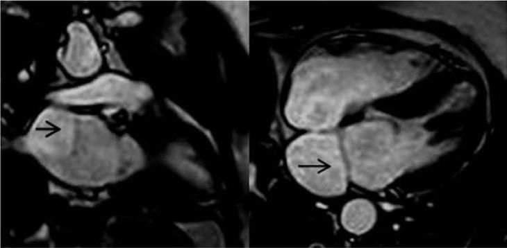
Establishing an accurate diagnosis of cor triatriatum requires a systematic imaging pathway to visualize the membrane, evaluate its restrictive gradient, and assess pulmonary vascular pressures. Transthoracic Echocardiography (TTE) serves as the primary non-invasive diagnostic modality, directly visualizing the fibromuscular membrane traversing the left atrium and identifying the proximal and distal sub-chambers. Color Doppler and continuous-wave Doppler echocardiography quantify the peak and mean pressure gradients across the membrane fenestrations, while assessing right ventricular function and pulmonary artery pressures. Transesophageal Echocardiography (TEE) provides superior structural detail in adults and complex cases, mapping the precise location, thickness, and fenestrations of the membrane relative to the pulmonary vein orifices and the left atrial appendage. Computed Tomography (CT) Angiography and Cardiac Magnetic Resonance (CMR) imaging offer three-dimensional anatomical reconstruction, allowing surgical teams to delineate pulmonary venous drainage patterns and plan operative access. Invasive cardiac catheterization is reserved for cases requiring direct pressure measurements or when non-invasive imaging remains inconclusive.
Therapeutic Interventions and Surgical Resection Strategies
Definitive management for symptomatic or hemodynamically restrictive cor triatriatum is strictly surgical. In critical neonatal presentations with severe obstruction and acute pulmonary edema, initial medical stabilization includes oxygen administration, diuretic therapy to reduce pulmonary congestion, and inotropic support if right ventricular failure is present. Once stabilized, open surgical repair using median sternotomy and cardiopulmonary bypass is performed. The surgical procedure involves opening the left atrium, carefully identifying the intra-atrial fibromuscular membrane, and excising it completely to unify the proximal and distal atrial chambers into a single, unobstructed cavity. Any co-existing atrial septal defects or anomalous venous connections are repaired simultaneously. Following complete surgical resection, the hemodynamic obstruction is permanently eliminated, pulmonary venous pressures rapidly normalize, and long-term prognosis is excellent, with minimal risk of late recurrence or structural complications.
Conclusion
Cor Triatriatum is a rare congenital cardiac defect characterized by an intra-atrial fibromuscular membrane that divides the atrium into two functional compartments, creating a mechanical obstacle to venous return. Understanding how restrictivemembrane fenestrations elevate proximal atrial pressures, induce postcapillary pulmonary hypertension, and strain the right heart clarifies the progression from infantile respiratory distress to adult exertional dyspnea and arrhythmias. Utilizing transthoracic and transesophageal echocardiography alongside advanced CT angiography enables clinical teams to accurately map the membrane architecture and quantify the pressure gradient. Through timely surgical resection under cardiopulmonary bypass, healthcare professionals can completely eliminate the atrial obstruction, restore normal pulmonary venous transit, and ensure excellent long-term cardiovascular health and life expectancy for affected individuals.