What Is Arrhythmogenic Mitral Valve Prolapse and How Can It Affect Heart Rhythm?
The mitral valve complex is a sophisticated anatomical structure designed to maintain unidirectional blood flow from the left atrium to the left ventricle during ventricular diastole and prevent retrograde flow during systole. This complex consists of two valve leaflets (anterior and posterior), the fibrous mitral annulus, chordae tendineae, and papillary muscles anchored within the left ventricular myocardium. Classic Mitral Valve Prolapse (MVP) is characterized by myxomatous degeneration of the valve leaflets, resulting in the displacement of one or both leaflets by more than two millimeters beyond the plane of the mitral annulus into the left atrium during systole. While classic MVP is generally considered a benign condition affecting a notable portion of the general population, a distinct high-risk subgroup exhibits severe ventricular dysrhythmias and an elevated risk of sudden cardiac death (SCD). This specific clinical entity, designated as Arrhythmogenic Mitral Valve Prolapse (AMVP), represents a mechanical and electrophysiological disorder where continuous mechanical traction exerted by prolapsing leaflets triggers localized myocardial remodeling and life-threatening electrical instability.
Anatomical Substrates and Mitral Annular Disjunction Mechanics
The primary structural driver of arrhythmogenesis in Arrhythmogenic Mitral Valve Prolapse involves abnormal mechanical coupling between the valve apparatus and the underlying left ventricular myocardium. A key anatomical hallmark frequently present in these patients is Mitral Annular Disjunction (MAD). Mitral annular disjunction is a structural deformity defined by a distinct spatial separation between the junction of the posterior mitral valve leaflet insertion point and the basal left ventricular free wall myocardium. During ventricular systole, the presence of MAD causes the mitral annulus to detach and hyper-pivot, leading to paradoxical systolic expansion and curling of the basal inferolateral ventricular wall. As the left ventricle contracts against high pressure, the prolapsing valve leaflets exert excessive systolic traction through the chordae tendineae directly onto the attached papillary muscles and the adjacent basal inferolateral myocardium.
Mechanisms of Myocardial Stretch, Fibrosis, and Arrhythmogenesis
The mechanical hyper-traction experienced by the papillary muscles and basal inferolateral ventricular wall initiates a cascade of localized tissue trauma and electrophysiological instability. Repetitive, high-force mechanical stretching during each cardiac cycle induces localized ischemia, cellular injury, and reactive interstitial fibrosis within the body of the papillary muscles and the adjacent subendocardial myocardium. On an electrophysiological level, mechanical stretch directly alters cardiomyocyte membrane potentials through stretch-activated ion channels, generating delayed afterdepolarizations (DADs) and triggering premature ventricular contractions (PVCs). Over time, localized dense myocardial fibrosis creates heterogenous conduction zones characterized by slow, fragmented electrical propagation. These fibrotic tracks form anatomical substrates capable of sustaining re-entrant electrical circuits. When triggered by mechanically induced premature ventricular beats, these re-entrant circuits can rapidly degenerate into sustained monomorphic or polymorphic ventricular tachycardia and fatal ventricular fibrillation.
Clinical Manifestations, Electrocardiographic Patterns, and Red Flags
Patients presenting with Arrhythmogenic Mitral Valve Prolapse exhibit a clinical spectrum ranging from mild symptomatic palpitations to sudden cardiac arrest. Common presenting symptoms include lightheadedness, exertional or non-exertional chest pain, presyncope, and syncopal episodes resulting from transient runs of non-sustained or sustained ventricular tachycardia. Physical examination classically reveals a mid-systolic click followed by a late systolic murmur best heard at the cardiac apex. Surface electrocardiography (ECG) provides vital clues regarding arrhythmic risk; characteristic electrophysiological red flags include T-wave inversions or ST-segment depressions in the inferior vascular leads (leads II, III, and aVF), reflecting localized metabolic stress and structural remodeling within the inferolateral myocardium. Continuous Holter monitoring frequently captures a high burden of complex ventricular ectopy, including frequent multiform PVCs, ventricular doublets, triplets, and non-sustained ventricular tachycardia (NSVT) originating from the papillary muscles, fascicular conduction pathways, or the mitral annulus.
Diagnostic Protocols, Advanced Cardiac Imaging, and Risk Stratification
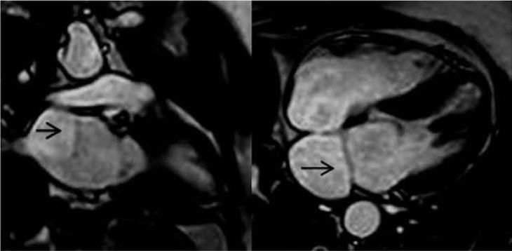
Establishing a diagnosis of Arrhythmogenic Mitral Valve Prolapse and stratifying individual arrhythmic risk requires a comprehensive multimodal evaluation. Transthoracic (TTE) and Transesophageal (TEE) Echocardiography provide precise anatomical visualization of leaflet prolapse, myxomatous thickness (exceeding five millimeters), the severity of mitral regurgitation, and the presence and distance of mitral annular disjunction during end-systole. Cardiac Magnetic Resonance (CMR) imaging serves as the indispensable gold standard for tissue characterization and risk stratification. Contrast-enhanced CMR using Late Gadolinium Enhancement (LGE) techniques allows clinicians to identify and visualize microvascular replacement fibrosis specifically within the papillary muscles, basal inferolateral wall, and mitral annulus. The presence of LGE in these regions correlates strongly with ventricular arrhythmia severity and heightened risk of sudden cardiac collapse. Additional risk factors elevating clinical concern include a history of unexplained syncope, female sex, bileaflet prolapse involvement, high PVC burden, and documented runs of complex non-sustained ventricular tachycardia.
Therapeutic Interventions and Electrophysiological Management
Managing Arrhythmogenic Mitral Valve Prolapse focuses on suppressing symptomatic dysrhythmias, preventing sudden cardiac death, and addressing significant valvular dysfunction. Medical therapy represents the first-line approach for symptomatic patients with non-sustained ventricular dysrhythmias; cardioselective beta-blockers (such as metoprolol or bisoprolol) reduce sympathetic drive, lower heart rate, and decrease mechanical stretch forces on the papillary muscles. Antiarrhythmic drugs like sotalol or amiodarone may be considered for refractory ventricular ectopy. In patients with drug-resistant, highly symptomatic ventricular arrhythmias originating from a focal source (such as a specific papillary muscle or the mitral ring), invasive catheter ablation utilizing electroanatomical mapping can successfully eliminate the arrhythmogenic focus. When severe mitral regurgitation is present alongside structural prolapse, surgical mitral valve repair or replacement reduces mechanical traction and restores normal intracardiac hemodynamics.
Conclusion
Arrhythmogenic Mitral Valve Prolapse is a distinct clinical variant of mitral valve disease defined by mechanical hyper-traction, localized myocardial fibrosis, and life-threatening ventricular dysrhythmias. Understanding how mitral annular disjunctionand leaflet prolapse exert continuous mechanical strain on papillary muscles and the inferolateral left ventricular wall clarifies the transition from mechanical stretch to fibrotic re-entrant circuits and fatal electrical instability. Utilizing high-resolution echocardiography, Holter monitoring, and late gadolinium enhancementcardiac MRI enables care teams to identify structural disjunction, map myocardial fibrosis, and stratify individual arrhythmic risk effectively. Through targeted beta-blocker therapy, catheter ablation of arrhythmogenic foci, timely ICD implantation, and surgical valve repair when indicated, healthcare professionals can effectively suppress ventricular arrhythmias, prevent sudden cardiac death, and optimize long-term cardiovascular outcomes for affected individuals.