Understanding Common Disorders of the Salivary Glands
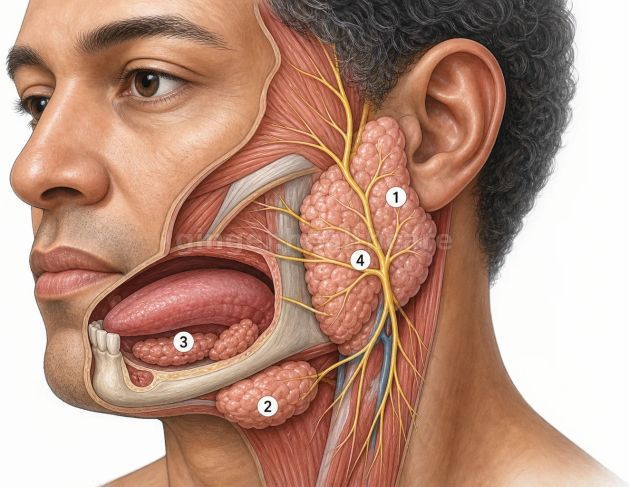
The human oral cavity relies upon a continuous, well-regulated supply of saliva to maintain oral hygiene, facilitate mastication, initiate chemical digestion, and protect mucosal tissues from pathogenic microbial invasion. This essential biological fluid is produced and secreted by three major pairs of salivary glands—the parotid, submandibular, and sublingual glands—alongside hundreds of minor salivary glands distributed throughout the mucous membranes of the lips, cheeks, and palate. The parotid glands, situated just in front of the ears, secrete primarily serous, watery fluid rich in digestive enzymes through Stensen's ducts. The submandibular glands, located beneath the jawbone, produce a mixed serous and mucous secretion delivered via Wharton's ducts, while the sublingual glands secrete thick, viscous mucus. When inflammatory processes, mechanical obstructions, autoimmune attacks, or neoplastic growths disrupt the normal architecture or secretory function of these glands, individuals develop a diverse spectrum of medical conditions collectively known as salivary gland disorders.
Sialolithiasis: The Pathophysiology of Salivary Duct Stones
One of the most frequent mechanical complications affecting the salivary system is sialolithiasis, a condition characterized by the formation of calcified concretions, or salivary stones, within the ductal networks or glandular parenchyma. These mineral deposits form primarily within the submandibular gland and its long, upward-sloping Wharton's duct, an anatomical pathway that predisposes secretions to stagnation and calcium precipitation. Salivary stones are composed predominantly of calcium phosphate and calcium carbonate, bound together by organic matrices of salivary proteins and cellular debris. When a growing stone completely or partially blocks the main excretory duct, saliva is prevented from flowing freely into the oral cavity. This obstruction causes a sudden buildup of fluid pressure behind the blockage, resulting in painful, recurrent swelling of the affected gland during meals when salivary stimulation peaks, frequently followed by gradual drainage once eating concludes.
Sialadenitis: Microbial Infections and Inflammatory Responses
When salivary flow is obstructed by stones, strictures, or dehydration, or when systemic illness suppresses salivary production, the stagnant environment within the ductal system becomes highly vulnerable to ascending bacterial colonization, precipitating acute or chronic sialadenitis. Acute suppurative sialadenitis is characterized by painful bacterial infection of the gland, most frequently incited by oral pathogens like Staphylococcus aureus ascending through the ductal opening. Patients experience rapid glandular enlargement, intense throbbing pain, localized erythema, and systemic manifestations such as high fever and leukocytosis, occasionally resulting in localized abscess formation that requires urgent medical drainage. Chronic sialadenitis, by contrast, develops from recurrent inflammatory insults or persistent low-grade ductal obstruction, leading to progressive destruction of acinar cells, extensive fibrosis, and permanent loss of secretory function.
Sialadenosis and Non-Inflammatory Glandular Enlargement
Beyond acute infections and mechanical obstructions, the salivary glands can undergo diffuse, non-inflammatory enlargement driven by systemic metabolic or endocrine disturbances, a condition designated as sialadenosis. This non-neoplastic disorder typically affects the parotid glands symmetrically, presenting as a soft, painless, and persistent swelling of the cheeks without signs of acute inflammation, redness, or purulent ductal discharge. Sialadenosis is frequently linked to underlying systemic conditions such as chronic alcoholism, diabetes mellitus, severe protein malnutrition, hormonal imbalances, and the adverse effects of certain prescription medications that alter autonomic control over salivary secretion. Histological evaluations reveal that this condition stems from abnormal hypertrophy of the acinar cells, characterized by an abnormal accumulation of secretory zymogen granules within the cytoplasm, alongside interstitial edema and fatty infiltration of the glandular tissue.
Autoimmune Pathologies: Sjögren's Syndrome and Xerostomia
Salivary glands are also primary targets for destructive autoimmune disorders, with Sjögren's syndrome standing as the most prominent systemic autoimmune condition affecting exocrine function. Sjögren's syndrome is characterized by chronic, progressive lymphocytic infiltration of the lacrimal and salivary glands, mediated by autoreactive T-lymphocytes and B-cells that gradually destroy the secretory acini and small ducts. This extensive glandular destruction leads to profound, chronic secretory failure, producing severe dry mouth, known medically as xerostomia, alongside dry eyes. The lack of protective saliva compromises oral health, leaving patients vulnerable to rampant dental caries, oral candidiasis, persistent mucosal trauma, and difficulty swallowing or speaking. Concurrently, patients suffer from persistent, uncomfortable swelling of the major salivary glands, particularly the parotid glands, reflecting the ongoing autoimmune inflammatory response within the tissue.
Neoplastic Tumors: Benign and Malignant Growths
The salivary glands are uniquely capable of developing a remarkably diverse array of benign and malignant tumors, accounting for a small percentage of all head and neck neoplasms. The majority of salivary gland tumors arise within the parotid glands, and approximately eighty percent of these parotid masses are benign, most commonly presenting as a slow-growing, painless pleomorphic adenoma. Pleomorphic adenomas feature a complex histological architecture containing epithelial and stromal elements, and while benign, they possess a potential for recurrence if incompletely excised. Conversely, smaller salivary glands—such as those in the submandibular region or hard palate—carry a significantly higher probability of harboring malignancy. Malignant salivary tumors include mucoepidermoid carcinoma, adenoid cystic carcinoma, and acinic cell carcinoma, which can invade local facial nerves, cause chronic pain, and metastasize regionally.
Diagnostic Evaluations and Clinical Investigations
Diagnosing disorders of the salivary glands requires a comprehensive clinical evaluation, beginning with a meticulous physical examination that involves palpating the glands and assessing ductal orifices for the expression of clear saliva versus purulent discharge. When structural pathology is suspected, clinicians utilize advanced imaging modalities. Ultrasonography serves as a non-invasive, highly effective initial tool to identify salivary stones, assess ductal dilation, and differentiate solid masses from fluid-filled cysts. Sialography—involves injecting radiopaque contrast dye directly into the ductal system—provides detailed fluoroscopic mapping of strictures and stone locations, though it has been largely superseded by magnetic resonance sialography and computed tomography scans. When tumors or persistent inflammatory masses are detected, fine-needle aspiration biopsy is frequently performed to obtain cellular samples for definitive histopathological analysis.
Management Strategies and Therapeutic Interventions
Therapeutic management of salivary gland disorders varies widely depending on the underlying etiology, ranging from conservative hydration and massage to surgical intervention. For obstructive conditions like sialolithiasis, smaller stones can often be dislodged conservatively through glandular massage, tart beverages to stimulate salivary flow, and adequate hydration, whereas larger or impacted stones are managed using minimally invasive sialendoscopy or surgical removal. Bacterial sialadenitis requires targeted antibiotic therapy combined with warm compresses and sialogogues, while acute abscesses necessitate surgical incision and drainage. Autoimmune xerostomia focuses on palliative moisture replacements, meticulous oral hygiene, and systemic immunomodulatory medications. Finally, neoplasms and chronic, non-functional, chronically infected glands typically require surgical resection, executed with meticulous care to protect critical neighboring structures like the facial nerve.
Conclusion
Salivary gland disorders encompass a wide spectrum of mechanical, infectious, autoimmune, and neoplastic conditions that disrupt the vital production and delivery of saliva within the oral cavity. By examining how salivary stones cause painful ductal obstruction, understanding the mechanisms of bacterial sialadenitis and autoimmune Sjögren's syndrome, and recognizing the diagnostic value of ultrasonography and fine-needle aspiration biopsy, the clinical importance of these glands becomes evident. Through precise medical management, minimally invasive endoscopic techniques, and targeted surgical interventions, healthcare providers can alleviate symptoms, eradicate infections, preserve glandular function, and restore oral health for affected individuals.