What Is Anomalous Left Coronary Artery From the Pulmonary Artery (ALCAPA)?
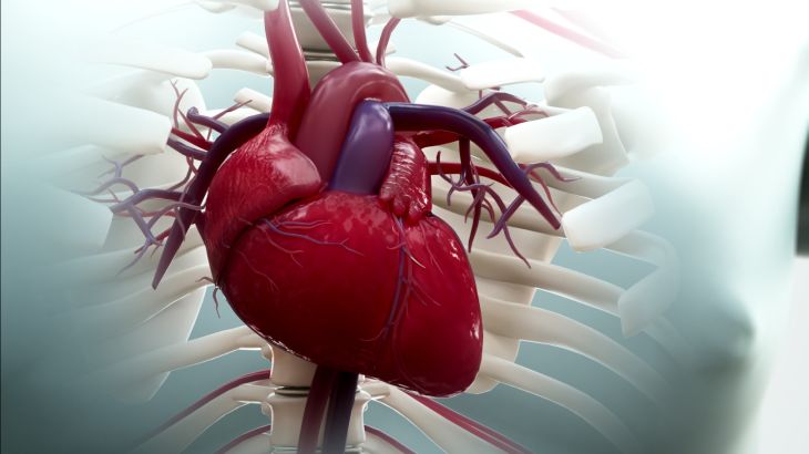
Normal cardiac function and cellular viability rely on an uninterrupted supply of oxygenated blood delivered via the coronary arterial system. Under normal anatomical conditions, the right coronary artery (RCA) and the left main coronary artery (LMCA)—which divides into the left anterior descending (LAD) and left circumflex (LCx) branches—originate from distinct sinuses of Valsalva within the root of the ascending aorta. Because aortic diastolic pressure is high, oxygenated blood flows smoothly into the coronary arterial network, nourishing the thick muscular walls of the left and right ventricles. Anomalous Left Coronary Artery from the Pulmonary Artery (ALCAPA), historically designated as Bland-White-Garland syndrome, is a rare, severe congenital cardiac malformation in which the left main coronary artery arises abnormally from the low-pressure pulmonary artery rather than the high-pressure aorta. This anatomical displacement fundamentally alters coronary perfusion dynamics, subjecting the infant myocardium to severe chronic ischemia, left ventricular dysfunction, mitral regurgitation, and early mortality if left uncorrected.
Embryological Etiology and Morphological Classifications
The developmental origin of ALCAPA stems from maldevelopment during early cardiac septation, specifically around the fifth to eighth week of gestation. During normal embryogenesis, the truncus arteriosus divides into the ascending aorta and the pulmonary trunk, while endothelial buds emerge from the aortic sinuses to form the primitive coronary arteries. Failure of normal division, abnormal placement of the coronary endothelial buds, or erroneous fusion of the left coronary bud with the pulmonary trunk sinus gives rise to ALCAPA. Morphologically, the anomalous left coronary artery most frequently arises from the posterior or left sinus of the main pulmonary artery near the bifurcation, though origins from the proximal right or left pulmonary artery branches can occur.
Pathophysiology of Coronary Steal and Ischemic Transition
The hemodynamic consequences of ALCAPA undergo a dramatic transition during the early neonatal period. In utero, fetal pulmonary vascular resistance is high, and blood pressure within the main pulmonary artery equals or exceeds systemic aortic pressure. Consequently, blood flows forward from the pulmonary artery into the anomalous left coronary artery, keeping the fetal left ventricular myocardium adequately perfused, albeit with relatively deoxygenated fetal blood. Following birth, as the infant's lungs inflate, pulmonary vascular resistance drops rapidly over the first few weeks of life, causing pulmonary artery pressure to fall. This drop in downstream pressure creates a profound hemodynamic dilemma. As pulmonary arterial pressure decreases below systemic levels, perfusion pressure within the anomalous left coronary artery drops, causing myocardial hypoperfusion and localized ischemia across the anterolateral left ventricular wall.
As the infant matures further, extensive collateral arterial vessels form between the high-pressure, normally attached right coronary artery and the low-pressure anomalous left coronary tree. Once these inter-coronary collaterals develop, blood supplied by the right coronary artery flows into the left coronary system. However, because the anomalous left coronary artery connects directly to the low-pressure pulmonary artery, blood takes the path of least resistance: it flows retrograde through the anomalous left coronary artery and dumps directly into the pulmonary artery. This continuous retrograde shunting is known as the "coronary steal phenomenon." Rather than perfusing the left ventricular myocardium, oxygenated blood from the right coronary artery bypasses the capillary beds and drains into the pulmonary circulation, exacerbating left ventricular ischemia, driving extensive subendocardial infarction, and triggering rapid cardiac dilation.
Clinical Spectrum: Infantile versus Adult Phenotypes
ALCAPA presents primarily across two distinct clinical phenotypes dictated by the speed and extent of collateral vascular network development. The infantile presentation represents the vast majority (roughly eighty to ninety percent) of cases. In these infants, inter-coronary collaterals are sparse or inadequate. As pulmonary vascular resistance drops during the first two months of life, severe myocardial ischemia ensues. Infants exhibit characteristic episodes of anginal distress, manifested by crying, irritability, diaphoresis, pallor, and tachypnea during feeding or exertion. As left ventricular systolic dysfunction worsens, infants develop severe dilated cardiomyopathy, poor feeding, failure to thrive, hepatomegaly, and overt congestive heart failure. Secondary ischemic dysfunction or papillary muscle infarction frequently induces severe mitral regurgitation. Without surgical intervention, up to ninety percent of affected infants die within the first year of life.
Conversely, a small subset of individuals possesses extensive, highly developed inter-coronary collateral networks that sustain borderline left ventricular perfusion into late childhood or adulthood. Patients with adult-type ALCAPA may remain relatively asymptomatic for years, though they face chronic subendocardial ischemia, localized myocardial scarring, and high risks of exertion-induced malignant ventricular arrhythmias, syncope, and sudden cardiac death.
Diagnostic Protocols and Cardiovascular Imaging
Establishing a definitive diagnosis of ALCAPA requires a high index of suspicion supported by non-invasive electrophysiological and cardiovascular imaging modalities. Electrocardiography (ECG) provides crucial diagnostic clues, characteristically showing deep Q waves in leads I, aVL, and V4 through V6, alongside ST-segment changes and T-wave inversions indicative of anterolateral myocardial infarction. Transthoracic Echocardiography (TTE) serves as the primary diagnostic tool. Color flow Doppler demonstrates abnormal retrograde blood flow from the anomalous left coronary artery entering the main pulmonary artery, dilated right coronary artery architecture, extensive inter-coronary collateral flow within the ventricular septum, and significant left ventricular dilation with reduced ejection fraction.
Therapeutic Interventions and Surgical Revascularization
The management of ALCAPA is strictly surgical, with prompt operative repair indicated upon diagnosis to establish a dual-coronary arterial system and restore two-vesselsystemic perfusion. Preoperative medical management focuses on stabilizing acute heart failure using diuretics, afterload-reducing vasodilators, and positive inotropic agents. The surgical procedure of choice in neonates and infants is Direct Coronary Translocation (reimplantation). Performed under cardiopulmonary bypass, the anomalous left coronary artery is excised with a surrounding button of pulmonary artery wall, mobilized, and directly reimplanted into the ascending aorta.
In scenarios where the anomalous origin is anatomically distant from the aorta, alternative surgical techniques are utilized. The Takeuchi procedure constructs an intra-pulmonary tunnel using a flap ofpulmonary artery wall to direct blood from a created aortopulmonary window to the orifice of the anomalous coronary artery. In adult presentations with heavily calcified or immovable vessels, coronary artery bypass grafting (CABG) using an internal mammary artery graft paired with ligation of the anomalous origin at the pulmonary artery prevents continuous retrograde steal. Following surgical revascularization, left ventricular geometry and systolic function often demonstrate remarkable recovery due to the high remodeling capacity of the pediatric myocardium.
Conclusion
ALCAPA is a severe congenital coronary anomaly defined by the anomalous origin of the left coronary artery from the pulmonary artery, precipitating severe myocardial ischemia, coronary steal, and dilated cardiomyopathy. Understanding how postnatal drops in pulmonary vascular resistance shift perfusion dynamics into retrograde collateral shunting clarifies the mechanics behind infantile heart failure, mitral insufficiency, and sudden cardiac death. Utilizing surface electrocardiography, color Doppler echocardiography, and multi-slice CT angiography enables clinical teams to establish an accurate diagnosis and map coronary anatomy effectively. Through timely surgical revascularization—specifically direct aortic reimplantation—healthcare professionals can eliminate coronary steal, restore dual-vessel myocardial perfusion, and facilitate complete recovery of left ventricular structure and function.