How Could 3D Ultrasound Imaging Improve the Detection of Certain Medical Conditions?
Medical imaging helps doctors look inside the body without performing surgery. Ultrasound is one of the most commonly used imaging methods because it uses sound waves instead of radiation to create images of internal structures.
Traditional ultrasound usually produces flat, two-dimensional images. However, 3D ultrasound imaging can create three-dimensional views of organs, tissues, and other body structures. These detailed views may help doctors understand certain medical conditions more clearly.
What Is 3D Ultrasound Imaging?
3D ultrasound imaging is a medical imaging technique that uses high-frequency sound waves to create three-dimensional pictures of structures inside the body.
Unlike conventional 2D ultrasound, which displays individual flat images, 3D ultrasound combines information from multiple imaging angles to build a more complete view.
Doctors can rotate or examine the resulting image from different directions. This may make it easier to understand the shape, size, and position of certain structures.
The technology is used in areas such as pregnancy care, heart examinations, and the assessment of selected organs and tissues.
How Does 3D Ultrasound Work?
The process uses sound waves and specialized computer software to create detailed images.
Sending Sound Waves Into the Body
A healthcare professional places an ultrasound probe against the skin, usually with a special gel to improve contact.
The probe sends high-frequency sound waves into the body. These waves travel through different tissues and reflect back toward the probe.
Collecting Image Information
The ultrasound system detects the returning echoes. Different tissues reflect sound waves in different ways, allowing the machine to collect information about internal structures.
Creating a 3D Image
The system combines information from multiple image sections to create a three-dimensional view.
Doctors can then examine the image from different angles, depending on the capabilities of the equipment.
Reviewing the Results
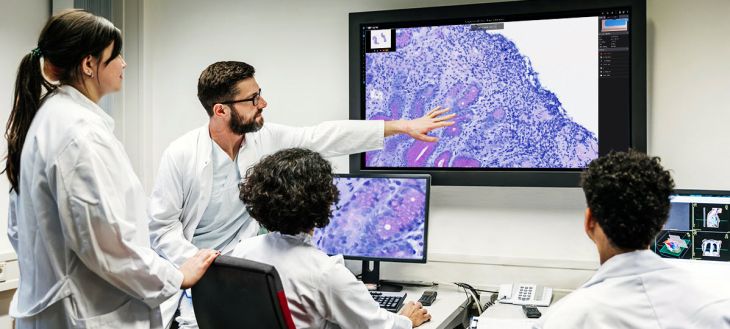
A trained healthcare professional studies the images to look for unusual structures, changes, or other signs that may require further investigation.
Which Medical Conditions Could Benefit From 3D Ultrasound?
3D ultrasound may help doctors examine certain conditions by providing a clearer view of anatomy. Its usefulness depends on the organ being examined and the quality of the images.
Pregnancy and Fetal Development
One of the familiar uses of 3D ultrasound is during pregnancy. It can create detailed images of a developing baby and show certain external features more clearly than standard 2D imaging.
Doctors may also use specialized ultrasound techniques to assess selected fetal structures, including the face and limbs.
However, 3D ultrasound does not replace routine pregnancy scans. Standard ultrasound remains important for checking fetal growth, position, and other medical measurements.
Heart Conditions
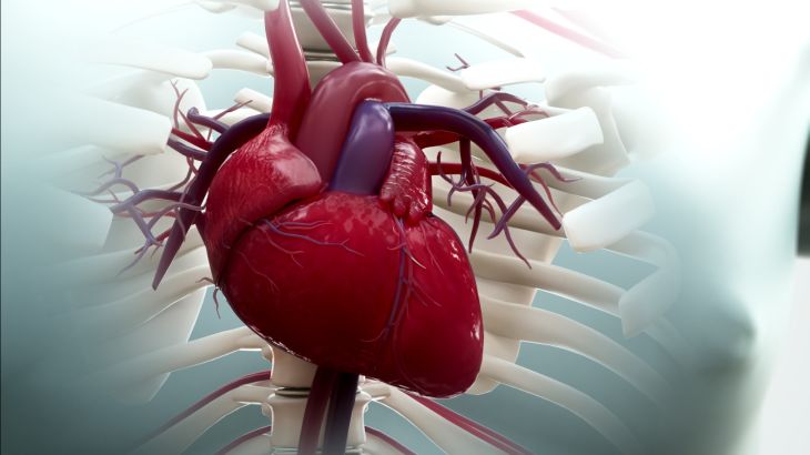
Three-dimensional echocardiography uses ultrasound to create detailed views of the heart.
It can help doctors examine the shape and movement of heart chambers and assess certain valve problems. It may also provide useful information about how the heart pumps blood.
This technique can be particularly valuable when doctors need to understand complex heart structures before planning treatment.
Blood Vessel Problems
Certain ultrasound systems can combine 3D imaging with Doppler technology, which evaluates blood movement.
This may help specialists examine the shape of blood vessels and assess blood flow in selected areas. Such information can be useful when investigating certain vascular abnormalities.
Organ Abnormalities
Researchers and clinicians are exploring 3D ultrasound for examining organs such as the liver, kidneys, and other structures.
Three-dimensional views may help doctors understand the location and shape of certain abnormalities. However, not every organ or condition can be assessed accurately with this method.
Musculoskeletal Conditions
Musculoskeletal ultrasound examines muscles, tendons, ligaments, and joints.
Three-dimensional imaging may help create a more complete view of selected injuries or structural changes. It could also assist specialists in understanding tissue damage and planning certain procedures.
What Are the Benefits of 3D Ultrasound Imaging?
Three-dimensional ultrasound offers several potential advantages in medical imaging.
More detailed views: It creates three-dimensional images that can make certain structures easier to understand.
Multiple viewing angles: Doctors can examine an image from different directions.
No ionizing radiation: Ultrasound uses sound waves rather than X-rays or other ionizing radiation.
Real-time assessment: Some ultrasound systems can capture movement and changes while an examination is taking place.
Treatment planning: Detailed images may help doctors prepare for selected medical procedures.
Non-invasive examination: Ultrasound generally does not require surgery or injections to create images.
These advantages depend on the type of examination and the condition being investigated.
What Are the Limitations of 3D Ultrasound?
Although 3D ultrasound can provide useful information, it has some limitations.
Image quality: The quality of an image may be affected by body position, movement, tissue depth, and other factors.
Limited visibility: Ultrasound waves do not pass easily through air or bone. This can make certain body structures difficult to examine.
Specialized equipment: Not every hospital or clinic has advanced 3D ultrasound systems.
Operator experience: Accurate image collection and interpretation require trained healthcare professionals.
Not suitable for every condition: Some medical problems require MRI, CT scans, or other diagnostic methods to obtain the necessary information.
3D ultrasound should therefore be selected according to the patient's symptoms and medical needs rather than used for every examination.
FAQs
Is 3D ultrasound better than 2D ultrasound?
Not always. 3D ultrasound provides three-dimensional views, which may be useful for examining certain structures. However, 2D ultrasound remains important for many routine examinations and medical measurements.
Is 3D ultrasound safe?
Ultrasound does not use ionizing radiation. When performed by trained professionals for appropriate medical reasons, it is generally considered safe. Medical examinations should follow recommended scanning practices.
Can 3D ultrasound detect cancer?
It may help doctors examine the shape and structure of certain masses or abnormalities. However, ultrasound alone cannot confirm every type of cancer. Additional imaging or a biopsy may be required.
Does 3D ultrasound take longer than a normal scan?
The examination time depends on the body area being studied, the equipment, and the information doctors need. Some 3D imaging procedures may require additional scanning and image processing.
Conclusion
3D ultrasound imaging is an important development in medical imaging because it allows doctors to examine certain internal structures from multiple angles. It may improve the assessment of heart conditions, fetal development, blood vessels, organs, and musculoskeletal problems.
Although it offers detailed images without using ionizing radiation, it cannot replace every traditional imaging method. Its effectiveness depends on the condition, equipment, and experience of the healthcare professional.