How Is AI-Assisted Microscopy Advancing Medical Diagnostics?
Microscopes have helped doctors and scientists study cells, tissues, and tiny organisms for many years. They play an important role in identifying infections, examining tissue samples, and understanding different diseases.
Today, artificial intelligence (AI) is adding new capabilities to microscopy. AI-assisted microscopy combines high-quality microscope images with computer systems that can recognize patterns and analyze large amounts of visual information.
This technology could help medical professionals examine samples more efficiently and identify changes that may need closer attention. Although it is promising, AI still needs careful testing and expert review before its results can be relied on for medical decisions.
What Is AI-Assisted Microscopy?
AI-assisted microscopy uses artificial intelligence to analyze images captured through a microscope.
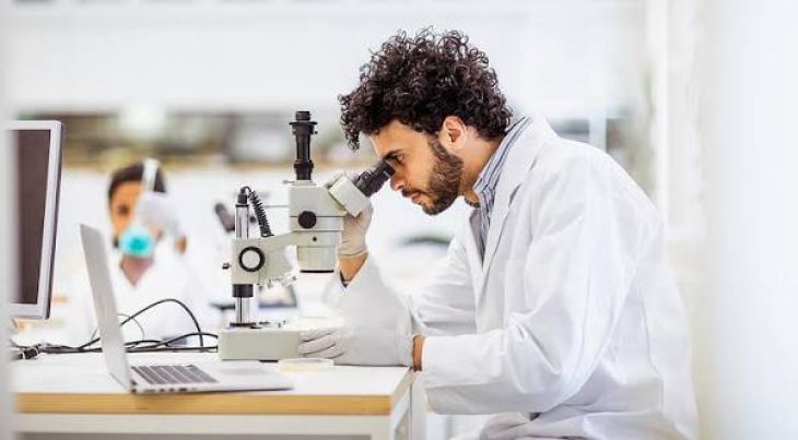
A traditional microscope allows a specialist to examine cells and other tiny structures. With AI-assisted microscopy, software can study these images, identify patterns, measure features, and highlight areas that may be unusual.
For example, an AI system may help count cells, identify certain microorganisms, or locate abnormal-looking cells in a tissue sample.
The technology is designed to assist laboratory professionals rather than replace their knowledge and judgment.
How Does AI-Assisted Microscopy Work?
The process usually begins with a microscope capturing an image of a sample.
The image is then analyzed by an AI model trained on relevant examples. Depending on the system, the software may identify cell shapes, measure sizes, count structures, or highlight patterns associated with particular conditions.
A laboratory specialist reviews the results and decides whether further testing is needed.
The quality of the results depends on several factors, including image quality, the data used to train the AI model, and the type of sample being examined.
How Can AI Microscopy Help Detect Diseases?
Some diseases cause changes in the appearance of cells, tissues, or microorganisms. These changes may be difficult to identify when samples contain thousands of tiny structures.
AI can examine many image features and highlight areas that deserve closer attention. This may help specialists find certain abnormalities more efficiently.
Researchers are studying AI-assisted microscopy for applications involving infections, blood disorders, tissue abnormalities, and other medical conditions.
However, the technology cannot detect every disease, and its performance varies depending on the specific application.
Can AI-Assisted Microscopy Improve Cancer Diagnosis?
Cancer diagnosis often involves examining tissue samples under a microscope. Pathologists look for changes in cell shape, tissue structure, and other features that may indicate cancer.
AI-assisted microscopy can help analyze digital images of these samples. Some systems are designed to highlight suspicious areas, count cells, or measure features that may help classify tumors.
This could make it easier for pathologists to review complex samples and focus on areas that need additional attention.
However, AI findings must be interpreted in context. A final cancer diagnosis may require expert examination, additional laboratory tests, and information about the patient's medical condition.
Could AI Help Identify Infections?
Microscopy is widely used in laboratories to examine samples for microorganisms and other signs of infection.
AI systems are being studied to help identify certain bacteria, parasites, and other microscopic structures. They may also help count cells or detect patterns that can guide further testing.
In some laboratory settings, these tools could reduce repetitive work and help specialists process samples more efficiently.
Still, the accuracy of AI-based identification depends on the organism, sample quality, and the system being used. Confirmatory testing may be necessary.
What Are the Benefits of AI-Assisted Microscopy?
AI-assisted microscopy could offer several benefits:
-
Faster analysis of certain samples
-
Automated counting of cells and microscopic structures
-
Identification of areas that need closer review
-
More consistent measurement of selected features
-
Assistance with large volumes of laboratory images
-
Better access to digital image sharing and research
These benefits depend on the quality of the technology and how well it fits into laboratory procedures.
What Are the Challenges?
AI-assisted microscopy also has limitations.
Poor image quality can affect results. AI models may perform differently when they analyze samples that differ from the data used during training.
Some systems may also produce false results, such as highlighting a harmless feature or missing an important abnormality.
Privacy is another concern because medical images may contain sensitive patient information. Laboratories must protect these records and follow appropriate data security practices.
Before an AI system is used for patient care, it needs suitable validation, quality checks, and monitoring.
What Is the Future of AI-Assisted Microscopy?
Researchers are working to make AI microscopy more accurate, reliable, and useful in real-world healthcare.
Future systems may combine microscope images with other medical information to help specialists understand complex conditions. AI could also support research by allowing scientists to measure and compare large numbers of cells more efficiently.
As the technology develops, its success will depend on reliable evidence, proper clinical testing, and cooperation between AI systems and trained medical professionals.
FAQs
Can AI-assisted microscopy diagnose diseases?
It can help identify patterns associated with certain diseases, but its role depends on the application. Medical professionals may need to confirm the findings with additional tests.
Can AI microscopy detect cancer cells?
Some AI systems can highlight cell and tissue features associated with cancer. Expert review remains important for diagnosis.
Does AI-assisted microscopy replace laboratory specialists?
No. It is generally designed to assist specialists by analyzing images and highlighting areas for review.
Is AI-assisted microscopy already being used?
Some AI-assisted microscopy tools are used in specific laboratory and research settings. Other applications are still being tested.
Can AI microscopy reduce laboratory errors?
It may help improve consistency in certain tasks, but AI can also make mistakes. Quality checks and human review remain essential.
Conclusion
AI-assisted microscopy is creating new ways to examine cells, tissues, and microorganisms. By analyzing microscope images and identifying patterns, it may help laboratory professionals work more efficiently and investigate diseases more accurately.
Although the technology offers promising opportunities in cancer research, infection testing, and other diagnostic fields, it is not a replacement for expert medical judgment. Careful testing and responsible use will be essential as AI microscopy becomes more widely adopted.