How Could Digital Pathology Help Laboratories Share and Analyze Tissue Samples More Efficiently?
Pathology plays an important role in modern healthcare. Doctors use tissue samples to identify diseases, understand abnormal cell changes, and make treatment decisions. Traditionally, pathologists examine these samples using glass slides and microscopes.
However, handling physical slides can take time, especially when samples need to be reviewed by specialists in different locations. Digital pathology is changing this process by allowing laboratories to turn tissue slides into high-resolution digital images.
These images can be stored, shared, and examined using computers. This technology could make laboratory collaboration easier, improve research, and help specialists analyze tissue samples more efficiently.
What Is Digital Pathology?
Digital pathology is the process of capturing, storing, managing, and analyzing pathology slides in digital form.
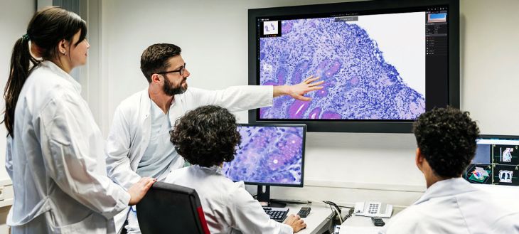
A special scanning device converts a glass tissue slide into a high-resolution image. This image can then be viewed on a computer screen using specialized software.
Instead of physically sending a slide to another laboratory, authorized specialists may be able to access its digital version through a secure system.
Digital pathology is used in several areas, including cancer diagnosis, disease research, medical education, and laboratory collaboration.
How Does Digital Pathology Work?
Digital pathology combines slide scanning, image storage, and computer-based analysis.
Preparing Tissue Samples
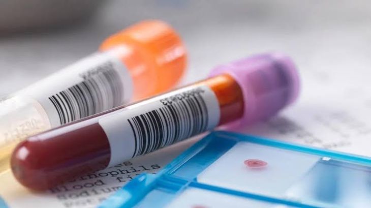
First, a tissue sample is collected from a patient through a biopsy or surgical procedure. The sample is processed in a laboratory and placed on a glass slide.
Special dyes are often used to make different cells and tissue structures easier to examine.
Scanning the Slides
A whole-slide imaging scanner captures the tissue at high resolution. It creates a digital image that can show cellular details similar to those seen through a microscope.
Storing Digital Images
The scanned images are stored on secure computer systems or specialized digital platforms. Laboratory staff can organize the files and retrieve them when needed.
Reviewing and Analyzing Images
Pathologists can examine digital slides by zooming in, moving across th image, and comparing different tissue areas.
Some platforms also include image analysis tools that can measure structures or highlight areas that may need closer examination.
How Could Digital Pathology Improve Laboratory Collaboration?
One of the biggest advantages of digital pathology is that it allows specialists to review the same tissue image without needing to handle the original glass slide.
Faster Sharing of Tissue Samples
Physical slides may need to be packed and transported between hospitals or laboratories. This can take time and create a risk of damage or loss.
Digital images can be shared through secure systems, allowing authorized specialists to review samples remotely.
Easier Expert Consultations
Some tissue samples are difficult to interpret. A local pathologist may want another specialist to review the findings.
Digital pathology makes it possible to share high-resolution images with experts in other hospitals or research centers. This can make second opinions and specialist consultations more convenient.
Better Communication Between Laboratories
Different laboratories may work together on complex cases or research projects. Digital images allow teams to discuss the same tissue areas and compare their observations more easily.
Improved Access to Specialist Knowledge
Smaller hospitals may not have specialists for every type of disease. Digital pathology could help connect these facilities with experts who have experience in rare cancers or unusual tissue conditions.
However, remote review depends on image quality, secure access, and appropriate clinical procedures.
Can Digital Pathology Help Detect Cancer?
Yes. Digital pathology can be used to examine tissue samples collected during cancer investigations.
Pathologists study cell shapes, tissue structures, and abnormal growth patterns to identify different types of cancer.
Digital slides allow specialists to zoom in on specific areas and compare tissue features. They may also use computer-assisted tools to measure tumor regions or identify areas that require further review.
Digital pathology can be particularly useful in cancer research because scientists can examine and compare large collections of tissue images.
However, digital images do not automatically confirm cancer. A qualified pathologist must interpret the findings alongside other clinical information and laboratory tests.
How Can Artificial Intelligence Improve Digital Pathology?
Artificial intelligence is becoming an important part of digital pathology research.
AI systems can be trained using large collections of labeled tissue images. Once trained and properly validated, they may help identify patterns that are difficult or time-consuming to measure manually.
Potential applications include:
Identifying suspicious areas: AI tools may highlight tissue regions that need closer examination.
Measuring tumor features: Software can help estimate tumor size, cell density, and other tissue characteristics.
Comparing tissue samples: Computer systems may help researchers identify similarities and differences between samples.
Organizing laboratory information: Automated tools can assist with image management and certain repetitive tasks.
Supporting research: AI can help scientists analyze large collections of digital slides more efficiently.
AI should be treated as an assisting tool rather than a replacement for trained pathologists. Its results require appropriate validation and professional review.
What Are the Benefits of Digital Pathology?
Digital pathology could offer several advantages for healthcare laboratories.
Convenient access: Authorized specialists can review digital images from different locations.
Reduced physical transportation: Laboratories may not need to send glass slides for every consultation.
Better image organization: Digital files can be stored and retrieved through organized systems.
Improved teaching: Medical students and trainees can study digital slides without needing access to physical microscope collections.
Research collaboration: Scientists can share and compare tissue images across institutions.
Efficient image analysis: Computer-based tools can assist with measurements and repetitive review tasks.
These benefits depend on suitable equipment, reliable software, and proper laboratory procedures.
What Challenges Does Digital Pathology Face?
Despite its advantages, digital pathology still has some limitations.
High equipment costs: Whole-slide scanners, storage systems, and specialized software can require significant investment.
Large file sizes: High-resolution tissue images need considerable storage space and reliable internet connections for sharing.
Data privacy: Patient information must be protected through secure storage, access controls, and appropriate data-sharing procedures.
Image quality: Poor scanning, incorrect slide preparation, or technical problems can affect the quality of digital images.
Training requirements: Pathologists and laboratory staff need training to use digital systems correctly.
Clinical validation: Digital workflows must be tested and validated before they can be used for routine diagnostic decisions.
What Is the Future of Digital Pathology?
The future of digital pathology may involve closer integration with artificial intelligence, cloud-based systems, and advanced medical research platforms.
Laboratories may increasingly use digital networks to share tissue images, request expert opinions, and study rare diseases. AI-assisted tools may also become more advanced, helping pathologists review images and organize complex information.
Another important development could be the creation of large digital tissue libraries. These collections may help researchers understand disease patterns and investigate new treatments.
However, digital pathology will need continued improvements in image quality, data security, affordability, and clinical validation.
FAQs
Is digital pathology the same as traditional pathology?
No. Both involve examining tissue samples, but digital pathology uses scanned images and computer software, while traditional pathology generally relies on glass slides and microscopes.
Can digital pathology help diagnose cancer?
Yes. Digital slides can be used to examine tissue structures and identify cancer-related changes. However, diagnosis still requires professional interpretation and appropriate laboratory procedures.
Can pathologists share tissue samples online?
They can share digital versions of tissue slides through secure platforms. This allows authorized specialists to review images without physically transporting the original slides.
Does digital pathology replace pathologists?
No. It provides digital tools that can make image review, collaboration, and research more convenient. Trained pathologists remain essential for interpreting results and making diagnostic decisions.
Conclusion
Digital pathology is changing how laboratories store, share, and examine tissue samples. By converting glass slides into high-resolution digital images, it allows specialists to collaborate remotely, review complex cases, and study tissue samples more conveniently.
The combination of digital imaging and artificial intelligence may further improve image analysis and medical research. Although challenges involving cost, data security, and clinical validation remain, continued progress could make pathology services more connected and efficient.