Understanding Common Disorders of the Bile Ducts and Their Warning Signs
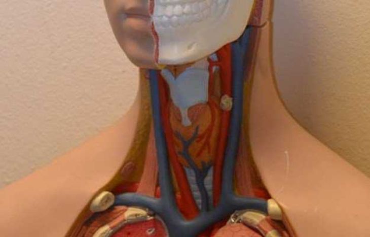
The intrahepatic and extrahepatic biliary transport system operates as an intricate physiological conduit network designed to deliver hepatic secretory fluid directly into the digestive tract. Continuously synthesized by functional liver parenchymal cells, bile constitutes a complex fluid mixture comprising conjugated bilirubin, bile acids, phospholipids, cholesterol, and inorganic ions. This liquid is indispensable for emulsifying dietary fats and aiding nutrient absorption, alongside facilitating the systemic elimination of endogenous metabolic byproducts. Branching intrahepatic canaliculi progressively merge into larger segmental channels, eventually uniting to form the principal left and right hepatic trunks, which converge outside the liver structure as the common hepatic conduit. The gallbladder joins this main pathway through the cystic duct, serving as a storage chamber to concentrate bile between digestive periods. Upon food ingestion, intestinal hormonal signals prompt gallbladder smooth muscle contraction, driving stored bile past the sphincter of Oddi into the duodenal chamber. Any physical interruption along these pathways compromises fluid movement, generating notable metabolic and systemic complications.
Mechanisms of Physical Blockage and Calculus Impaction
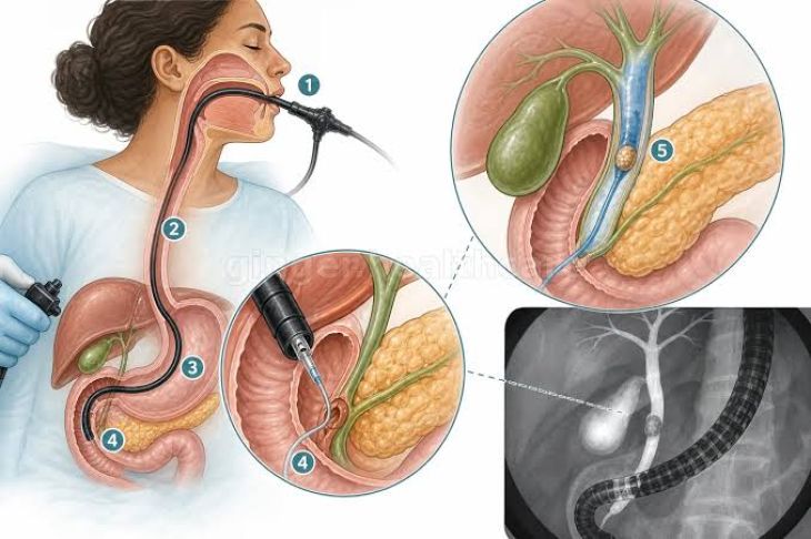
Choledocholithiasis remains a leading clinical pathology affecting the extrahepatic biliary conduits, characterized by the impaction of solid calculi within the primary common passage. These mineralized deposits frequently originate within the gallbladder reservoir—termed secondary ductal stones—and migrate into the main channel, though primary calculi can also form directly within stagnant ductal segments during periods of chronic fluid stasis or microbial growth. When a calculus becomes wedged within the narrow distal lumen or near the duodenal junction, physical fluid movement stops. This blockage rapidly elevates internal hydraulic pressure, driving conduit dilation, visceral capsule distention, and backpressure on upstream liver tissue. Prolonged fluid retention destabilizes the tight junctions connecting neighboring hepatocytes, redirecting conjugated bilirubin directly into the systemic vascular system.
Microbial Proliferation and Acute Infectious Complications
A major, life-threatening complication resulting from acute ductal occlusion is acute ascending cholangitis, a severe microbial invasion of the stagnant biliary network. When gallstones, fibrotic narrowings, or neoplastic growths obstruct natural fluid movement, static bile becomes a fertile environment for enteric microbes ascending from the duodenal lumen—most frequently Escherichia coli, Klebsiella pneumoniae, and Enterococcus species. As microbial density rises alongside escalating internal fluid pressure, the structural integrity of the mucosal lining deteriorates, allowing bacterial cells and endotoxic lipopolysaccharides to migrate into hepatic veins and systemic circulation. This systemic intrusion can rapidly trigger septic shock, systemic inflammatory response syndrome, and multi-organ failure if emergency biliary decompression is delayed.
Chronic Inflammatory Strictures, Autoimmunity, and Neoplastic Lesions
In addition to acute calculus impaction, disorders affecting the biliary conduits include chronic inflammatory, autoimmune, and neoplastic conditions. Primary Biliary Cholangitis targets smaller intrahepatic interlobular channels through granulomatous autoimmune destruction. Additionally, iatrogenic fibrotic strictures can develop following surgical trauma during gallbladder resection or as a consequence of persistent pancreatic inflammation. Within the neoplastic spectrum, cholangiocarcinoma—a malignant transformation of the ductal epithelium—presents as an insidious, slow-growing lesion that gradually occludes the conduit lumen, requiring early detection for potential surgical resectability.
Clinical Presentations and Systemic Warning Indicators
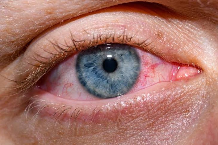
Recognizing early clinical warning signs is vital to preventing severe systemic morbidity associated with ductal occlusion. The classic clinical presentation signaling an obstructed biliary pathway includes scleral and cutaneous icterus, dark amber urine, and pale acholic stools. Because physical blockage prevents bile pigments from reaching the intestinal lumen, stercobilinogen production ceases, turning fecal matter a pale gray or clay color, while excess water-soluble conjugated bilirubin undergoes renal filtration, darkening the urine. Patients routinely report steady right upper quadrant abdominal discomfort radiating toward the shoulder region, accompanied by generalized pruritus caused by dermal bile salt deposition. In ascending cholangitis, the recognized presentation known as Charcot's triad comprises fever, jaundice, and right upper quadrant distress, which can progress to Reynolds' pentad with the emergence of hypotension and altered mental state.
Diagnostic Evaluations and Therapeutic Flow Restoration
Evaluating suspected ductal pathology requires combining biochemical serum testing with non-invasive and interventional imaging techniques. Biochemical panels typically reveal a classic cholestatic pattern, marked by pronounced elevations in circulating alkaline phosphatase, gamma-glutamyl transferase, and direct bilirubin. Transabdominal ultrasonography serves as the primary screening tool to detect internal ductal widening and identify cholelithiasis. For comprehensive, high-resolution mapping of the biliary architecture, Magnetic Resonance Cholangiopancreatography offers detailed visualization of strictures, anatomical variations, and radiolucent calculi without radiation exposure. When interventional clearance is required, Endoscopic Retrograde Cholangiopancreatography enables clinicians to access the duodenal papilla directly, perform a sphincterotomy, remove impacted calculi, or insert expandable endoprosthetic stents across strictured regions to restore unobstructed bile transit.
Conclusion
Disorders affecting the biliary conduits represent a significant category of gastrointestinal pathologies requiring rapid clinical identification and structured management. Understanding how calculus impaction, ascending microbial growth, autoimmune destruction, and malignant strictures impair normal fluid movement clarifies the physiological mechanics driving clinical symptoms. Identifying key warning signs—such as jaundice, acholic stools, dark urine, and right upper quadrant discomfort—enables timely diagnostic evaluation through advanced cross-sectional imaging and interventional endoscopy. Through prompt clinical management, medical teams can relieve internal conduit pressure, re-establish uninterrupted bile flow, and prevent life-threatening complications like ascending sepsis and chronic liver failure.