What Is Ménière’s Disease and How Can It Affect Hearing and Balance?
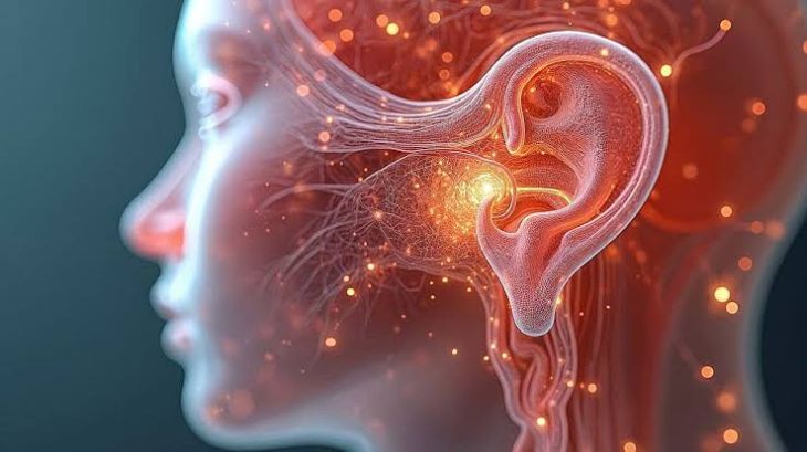
The inner ear houses two essential sensory systems integrated within a complex bony and membranous labyrinth: the cochlea, which processes acoustic vibrations, and the vestibular apparatus, which detects angular and linear acceleration to maintain spatial equilibrium. Both systems rely on two distinct fluid compartments to function correctly. Perilymph, a fluid rich in sodium, fills the bony labyrinth and surrounds the delicate membranous structures. Endolymph, a high-potassium fluid, fills the internal membranous space, bathing the hair cells of the organ of Corti and the vestibular cristae and maculae. Maintaining a strict chemical gradient and hydrostatic pressure differential between these two fluid spaces is vital for converting physical sound waves and head movements into neural signals. Ménière's disease represents a chronic, progressive disorder of the inner ear defined by the abnormal accumulation of endolymph, which disrupts delicate pressure balances, damages sensory receptor cells, and causes fluctuating hearing loss, episodic vertigo, tinnitus, and a sensation of aural fullness.
Pathophysiology of Endolymphatic Hydrops
The primary pathological feature underlying Ménière's disease is endolymphatic hydrops, an abnormal distension of the endolymphatic space within the membranous labyrinth. Endolymph is continuously produced by the stria vascularis within the cochlea and reabsorbed by the endolymphatic sac. Endolymphatic hydrops develops when there is an imbalance between fluid production and reabsorption, typically driven by impaired drainage through the endolymphatic duct. This fluid accumulation increases hydrostatic pressure within the membranous labyrinth, causing progressive distension of Reissner's membrane in the cochlea and the membranous walls of the saccule and utricle. As pressure elevates, micro-tears can occur in the thin membranous barrier, allowing potassium-rich endolymph to mix directly with perilymph. This sudden alteration in the extracellular fluid environment bathes the basilar membrane and vestibular afferent nerve fibers in high potassium concentrations, causing acute chemical depolarization, toxic neural blockade, and sudden vestibular and auditory dysfunction.
Mechanics of Vestibulo-Auditory Dysfunction and Episodic Vertigo
The periodic rupture and subsequent healing of the membranous labyrinth explain the classic episodic nature of Ménière's disease symptoms. When the membranous barrier breaks down, the sudden influx of potassium depolarizes the hair cells and primary afferent neurons of the vestibular nerve, triggering severe, spontaneous spinning vertigo. These vertiginous episodes are typically accompanied by spontaneous horizontal-torsional nystagmus, intense nausea, vomiting, diaphoresis, and severe postural instability. Concurrently, the elevated mechanical pressure within the cochlear duct alters the biomechanical properties of the basilar membrane. This mechanical stiffening predominantly affects the apex of the cochlea, where low-frequency sound vibrations are processed, producing the characteristic early symptom of low-frequency sensorineural hearing loss. Patients also experience continuous or fluctuating low-pitched tinnitus—often described as a roaring or ocean-like sound—alongside a sensation of pressure or fullness in the affected ear.
Disease Progression and Permanent Structural Damage
In the early stages of Ménière's disease, symptoms occur in discrete, paroxysmal attacks, with hearing thresholds and balance function returning toward baseline between episodes as the membranous tears heal and fluid homeostasis is temporarily restored. However, repeated episodes of endolymphatic hydrops, chemical toxicity, and chronic mechanical distension lead to irreversible structural degradation over time. Continuous micro-membrane ruptures and persistent metabolic stress cause progressive loss of sensory hair cells within the organ of Corti and vestibular maculae, accompanied by atrophy of the stria vascularis and fibrotic changes within the endolymphatic sac. Consequently, the temporary low-frequency hearing loss transitions into a permanent, flat, severe sensorineural hearing deficit across all frequencies, accompanied by profound speech discrimination impairment. Similarly, persistent vestibular destruction reduces acute vertigo frequency but leads to chronic, uncompensated vestibular hypofunction, persistent gait instability, and spatial disorientation.
Clinical Presentation and Diagnostic Evaluation Protocols
Diagnosing Ménière's disease relies on clinical diagnostic criteria established by international otolaryngological societies, combined with comprehensive neurotologic testing to exclude mimic conditions. Clinical criteria require at least two spontaneous episodes of vertigo lasting between twenty minutes and twelve hours, audiometrically documented low- to mid-frequency sensorineural hearing loss in the affected ear, and fluctuating auditory symptoms such as tinnitus or aural fullness. Serial pure-tone audiometry is essential to capture the low-frequency hearing loss pattern during or shortly after acute attacks. Electrocochleography (ECochG) can detect elevated summating-to-action potential ratios, reflecting endolymphatic pressure elevation. Magnetic Resonance Imaging (MRI) of the internal auditory canals with delayed post-contrast sequences allows direct visualization of endolymphatic hydrops while ruling out structural lesions such as vestibular schwannomas, demyelinating disease, or vascular loop compressions.
Therapeutic Management and Interventional Strategies
Because there is currently no cure for Ménière's disease, clinical management concentrates on reducing acute attack frequency, preserving long-term hearing, alleviating vertigo, and improving overall quality of life. Acute vertiginous attacks are managed symptomatically using vestibular suppressants (such as meclizine or benzodiazepines) and antiemetics to control nausea. Long-term prophylactic strategies aim to minimize endolymphatic fluid accumulation through lifestyle modifications, including strict dietary sodium restriction, caffeine and alcohol avoidance, and maintain adequate hydration. Pharmacological prophylaxis frequently utilizes oral diuretics (such as hydrochlorothiazide or acetazolamide) or betahistine to regulate endolymphatic pressure. For patients with refractory, disabling vertigo despite conservative therapy, interventional procedures are considered. Intratympanic corticosteroid injections offer non-destructive anti-inflammatory control, whereas intratympanic gentamicin ablation selectively eliminates vestibular hair cells to stop vertigo attacks. Surgical options—such as endolymphatic sac decompression, labyrinthectomy, or vestibular nerve section—are reserved for severe, intractable cases.
Conclusion
Ménière's disease is a complex inner ear disorder driven by endolymphatic hydrops, leading to structural distension, fluid barrier disruptions, and progressive sensorineural damage. Understanding how fluid pressure elevations and chemical mixing disrupt cochlear and vestibular hair cell mechanics clarifies the episodic vertigo, fluctuating hearing loss, tinnitus, and chronic balance instability experienced by affected individuals. Utilizing serial audiometry, electrocochleography, and advanced contrast-enhanced MRI enables multidisciplinary care teams to establish accurate diagnostic profiles and rule out central neurological pathology. Through structured dietary modifications, prophylactic diuretic therapy, intratympanic interventions, and targeted vestibular rehabilitation, healthcare professionalscan effectively control debilitating vertigo, minimize long-term auditory degradation, and support daily functional independence for patients living with this chronic inner ear condition.