Understanding Infective Endocarditis and Its Effects on the Heart
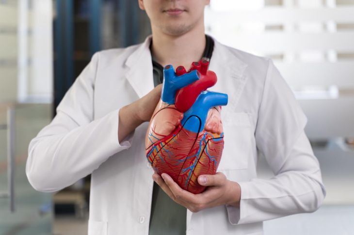
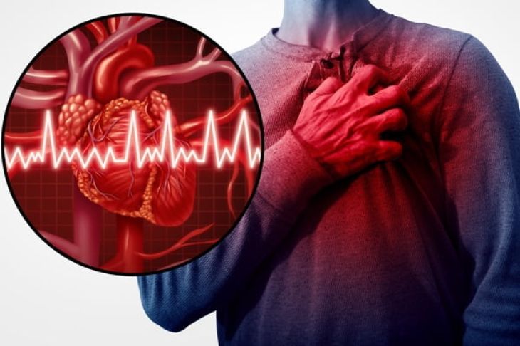
The human heart is lined internally by a smooth, glistening membrane known as the endocardium, which covers the heart chambers and forms the structural foundation of the cardiac valves. Under normal physiological conditions, this continuous endothelial layer maintains a non-thrombogenic surface that prevents blood cells from adhering and shields the deeper myocardial tissues from circulating pathogens. However, when structural abnormalities, congenital malformations, or degenerative changes alter the integrity of this lining, turbulent blood flow damages the endothelial cells, exposing the underlying extracellular matrix. This localized injury initiates the deposition of sterile platelets and fibrin, creating a receptive environment known as non-bacterial thrombotic endocarditis. If bacteria, fungi, or other microorganisms enter the bloodstream during transient bacteremia—originating from routine dental procedures, skin infections, or intravenous access—they can latch onto these sterile deposits. Once attached, these microbes proliferate rapidly, transforming the microenvironment into an active, destructive infection of the heart lining and valves, a serious condition known as infective endocarditis.
Pathological Formation of Cardiac Vegetations
The hallmark pathological feature of infective endocarditis is the development of cardiac vegetations, which are friable, mass-like structures composed of dense colonies of microorganisms tangled within a meshwork of fibrin, platelets, and inflammatory cells. These infected masses typically attach directly to the downstream surface of heart valves, most frequently targeting the mitral and aortic valves on the left side of the heart, though the tricuspid and pulmonary valves are commonly involved in individuals with intravenous drug use. As these vegetations grow, they disrupt the normal mechanical closure of the valve leaflets, causing acute valvular insufficiency or regurgitation. Furthermore, the localized inflammatory response triggered by the infection induces tissue necrosis, valve perforation, chordae tendineae rupture, and perivalvular abscess formation. This structural breakdown compromises the heart's hemodynamic integrity, rapidly precipitating acute heart failure as blood leaks backward through damaged valve openings.
Systemic Complications: Septic Emboli and Immune Phenomena
Beyond localized destruction within the cardiac chambers, infective endocarditis poses a severe threat to the entire body through the dissemination of septic emboli and immune-mediated phenomena. Because the vegetations are friable and constantly exposed to high-velocity arterial blood flow, tiny fragments composed of bacteria, platelets, and fibrin frequently break away from the main mass and travel downstream through the systemic circulation. These septic emboli lodge in various distant organ beds, causing localized tissue infarction, abscess formation, and severe organ damage. Common destinations include the cerebral circulation, leading to embolic strokes or brain abscesses; the spleen and kidneys, causing localized infarcts; and the pulmonary vasculature, particularly in right-sided endocarditis. Concurrently, the chronic presence of circulating antigens stimulates a robust immune response, resulting in the deposition of circulating immune complexes in small blood vessels. This immune activity generates characteristic clinical signs such as painful subcutaneous nodules on the fingers, painless hemorrhagic lesions on the palms, and tiny retinal hemorrhages known as Roth spots.
Etiological Agents: Bacterial and Fungal Pathogens
Infective endocarditis can be incited by a diverse array of microbial pathogens, though bacterial species account for the vast majority of documented clinical cases. Staphylococcus aureus has emerged as the most frequent and aggressive causative agent, capable of infecting both previously normal and structurally damaged heart valves, often resulting in rapid destruction and metastatic abscesses. Streptococci, including viridans group streptococci that naturally inhabit the oral cavity, are traditionally responsible for subacute presentations involving previously abnormal or rheumatic heart valves. Other notable bacterial contributors include the HACEK group of fastidious Gram-negative organisms, Enterococcus species acquired through gastrointestinal or urinary tracts, and opportunistic fungi like Candida species, which typically affect immunocompromised patients, prosthetic heart valves, or chronic intravenous catheter users. Identifying the exact offending pathogen through blood cultures is an absolute clinical prerequisite to guide targeted antimicrobial therapy.
Clinical Presentation and Symptom Variability
The clinical presentation of infective endocarditis is notoriously variable and deceptive, often mimicking numerous other systemic illnesses and earning it a reputation as a diagnostic chameleon. Subacute presentations develop insidiously over weeks to months, featuring vague, constitutional symptoms such as persistent low-grade fever, unexplained weight loss, night sweats, profound fatigue, generalized malaise, and new or changing heart murmurs heard during physical auscultation. Conversely, acute presentations driven by aggressive pathogens like Staphylococcus aureus strike with sudden, overwhelming severity, characterized by high spiking fevers, chills, rapid hemodynamic collapse, and acute signs of organ failure. Because microorganisms constantly shed from cardiac vegetations into the bloodstream, continuous bacteremia leads to persistent systemic inflammation, elevated inflammatory markers, and progressive physical debilitation if the underlying infection goes unrecognized.
Diagnostic Investigations: Modified Duke Criteria and Imaging
Diagnosing infective endocarditis relies on a comprehensive, systematic framework known as the modified Duke criteria, which integrates clinical findings, microbiological blood cultures, and advanced imaging results.
-
Microbiological Cultures: Obtaining multiple sets of blood cultures from separate venipuncture sites prior to initiating antibiotic therapy is mandatory to isolate the causative pathogen and determine its specific antimicrobial sensitivities.
-
Echocardiography: Transthoracic and transesophageal echocardiography serve as the primary imaging modalities for visualizing cardiac structures. Transesophageal echocardiography, in particular, offers exceptionally high resolution, allowing cardiologists to directly visualize oscillating intracardiac vegetations, measure valve perforation size, identify perivalvular abscesses, and assess the overall hemodynamic impact on ventricular function.
Antimicrobial Therapy and Surgical Intervention
Managing infective endocarditis requires prolonged, high-dose intravenous antimicrobial therapy designed to eradicate bacteria deeply embedded within avascular cardiac vegetations. Treatment regimens typically span four to six weeks, utilizing bactericidal antibiotics tailored precisely to the sensitivities of the isolated pathogen. Despite aggressive medical management, surgical intervention becomes necessary in a significant percentage of cases. Cardiothoracic surgeons must perform urgent or emergent valve repair or valve replacement when patients experience severe, intractable heart failure caused by valvular destruction; persistent systemic embolization despite adequate antimicrobial therapy; fungal endocarditis; or the formation of perivalvular abscesses that threaten the electrical conduction system of the heart.
Conclusion
Infective endocarditis is a severe, life-threatening infection of the heart's internal lining and valves, driven by microbial colonization that forms destructive vegetations and threatens systemic health through septic emboli. By exploring how damaged endothelial surfaces trap circulating bacteria, understanding the mechanisms of valvular destruction and embolic complications, and recognizing the diagnostic value of blood cultures and transesophageal echocardiography, the critical nature of this disease becomes clear. Through prolonged targeted intravenous antibiotic protocols and timely surgical valve interventions, healthcare providers can eradicate the infection, restore cardiac mechanics, and save patient lives.