Understanding Common Disorders of the Cornea and Their Effects on Vision
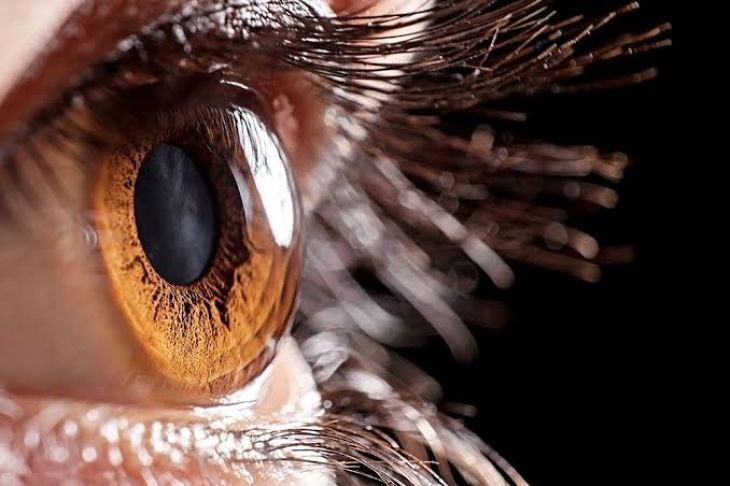
Visual acuity depends upon the optical clarity and anatomical curvature of the cornea, the transparent anterior structure of the eye. Functionally, the cornea serves as the primary refractive medium of the ocular system, accounting for approximately two-thirds of the total refractive power of the eye. Achieving crisp focus on the retina requires complete optical transparency, a precise spherical geometry, and a stable tear film layer. Structurally, the cornea consists of five primary layers: the non-keratinized anterior epithelium, Bowman's layer, the thick collagenous stroma, Descemet's membrane, and the innermost endothelial monolayer. Because the tissue is completely avascular to maximize light transmission, its metabolic demands are satisfied via aqueous humor diffusion and oxygen absorption from the atmospheric tear film. Structural disruption across any of these layers alters light refraction, scatters incoming visual signals, and leads to significant visual impairment.
Ectatic Disorders and Keratoconus Dynamics
Among structural corneal diseases, ectatic disorders represent a major cause of progressive visual distortion. Keratoconus is the most prevalent corneal ectasia, characterized by non-inflammatory, progressive thinning and weakening of the central and paracentral corneal stroma. At the cellular level, this condition involves enzymatic degradation of the extracellular matrix, loss of structural keratocytes, and mechanical slipping between adjacent collagen lamellae. As intraocular pressure acts upon this structurally weakened stroma, the central tissue bulges outward into an irregular, conical profile. This loss of normal spherical curvature generates severe irregular astigmatism and high-order optical aberrations that cannot be fully corrected with standard spectacles. Patients experience blurred vision, monocular polyopia, marked light sensitivity, and frequent shifts in refractive prescriptions.
Corneal Dystrophies and Endothelial Failure
Corneal dystrophies comprise a diverse group of hereditary, bilateral, non-inflammatory disorders that result in progressive accumulation of abnormal metabolic deposits within specific corneal layers. Fuchs Endothelial Dystrophy represents a classic, clinically significant variant affecting the innermost endothelial layer. Under normal physiological conditions, endothelial cells utilize active sodium-potassium ATPase pumps to continuously extract fluid from the stroma, maintaining a state of relative dehydration necessary for optical clarity. As fluid accumulates, the stroma and overlying epithelium undergo chronic edema, leading to corneal clouding, decreased contrast sensitivity, morning visual blurring, and painful epithelial bullae when fluid-filled blisters rupture.
Infectious Keratitis and Inflammatory Ulceration
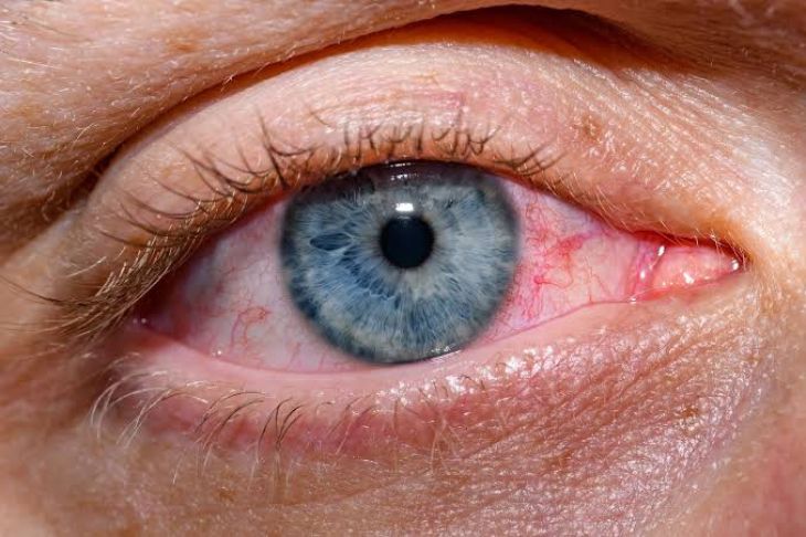
Infectious keratitis constitutes an acute, vision-threatening emergency where pathogens breach the protective anterior epithelial barrier to invade the corneal stroma. Bacterial, fungal, viral, and parasitic organisms represent the primary infectious etiologies. Bacterial keratitis often occurs following focal epithelial trauma or prolonged contact lens wear, commonly involving organisms such as Pseudomonas aeruginosa and Staphylococcus aureus. Microbial invasion triggers an intense host immune response, driving neutrophil infiltration, local tissue necrosis, and enzymatic breakdown of structural collagen. This destruction produces a localized corneal ulcer, dense stromal opacification, intense ocular pain, photophobia, and ciliary injection. If untreated, deep stromal ulceration can progress rapidly to corneal perforation, secondary intraocular infection, and irreversible scarring that permanently blocks visual transmission.
Corneal Trauma and Scarring Mechanisms
Mechanical trauma, chemical burns, and surgical complications present significant risks to corneal integrity and visual function. Transformed myofibroblasts deposit disorganized, dense collagen fibers that lack the regular spacing necessary to maintain optical transparency. This repair process forms opaque, white scar tissue known as a leucoma. Similarly, severe alkali chemical burns destroy limbal stem cells—the specialized progenitor cells situated at the corneal border responsible for epithelial regeneration. Loss of these stem cells leads to conjunctivalization, a pathological process where opaque vascularized conjunctival tissue grows over the clear corneal surface, causing profound visual loss.
Dry Eye Disease and Surface Epitheliopathy
Dry Eye Disease represents a multifactorial disorder of the ocular surface characterized by loss of tear film homeostasis and chronic local inflammation. The condition is broadly classified into aqueous-deficient dry eye, where the lacrimal glands produce inadequate fluid, and evaporative dry eye, driven by meibomian gland dysfunction and rapid tear breakdown. When the tear film thins or breaks down prematurely, the underlying corneal epithelium is exposed to environmental stress and hyperosmolar tear friction. This exposure induces punctate epithelial keratitis—microscopic erosions across the corneal surface. Patients report persistent burning, foreign body sensations, ocular fatigue, and episodic blurry vision that fluctuates with blinking. Over time, chronic surface inflammation damages corneal nerve fibers, compromising protective reflex blinking and accelerating surface breakdown.
Diagnostic Evaluations and Therapeutic Strategies
Accurate management of corneal pathology requires detailed structural and functional diagnostic assessments. Corneal topography and tomography map anterior and posterior surface curvature, measuring corneal thickness to identify early keratoconus. Anterior segment optical coherence tomography provides high-resolution cross-sectional images of individual corneal layers, quantifying stromal scars and fluid accumulation. Endothelial cell density and guttae formation are evaluated using specular microscopy. Treatment strategies range from conservative medical therapies to advanced surgical procedures. Dry eye and mild dystrophy cases are managed with lubricating agents, hypertonic saline drops, or anti-inflammatory medications. Severe ectasia is treated with corneal collagen cross-linking to structurally stabilize the stroma. When extensive stromal scarring, end-stage endothelial failure, or severe ulceration impairs vision, surgical options—such as deep anterior lamellar keratoplasty or endothelial keratoplasty—are performed to replace damaged tissue and restore visual function.
Conclusion
Corneal disorders encompass a broad spectrum of structural, hereditary, infectious, and degenerative conditions that disrupt optical clarity and refractive precision. Understanding how diseases like keratoconus, Fuchs dystrophy, infectious keratitis, and chronic dry eye impair corneal layers clarifies the mechanisms behind reduced visual acuity and surface irritation. Combining cross-sectional imaging with specialized topography allows clinical teams to accurately identify structural abnormalities and initiate targeted therapies.Through timely medical management, corneal collagen cross-linking, or targeted lamellar keratoplasty, clinicians can successfully halt disease progression, restore optical transparency, and preserve long-term visual health.