What Is Sleep Apnea and How Can It Affect the Brain?
Sleep is an essential biological state required for tissue repair, emotional regulation, and cognitive restoration. However, for millions of individuals worldwide, this nightly recovery process is repeatedly interrupted by a common yet serious medical condition known as sleep apnea. Sleep apnea is characterized by repetitive pauses in breathing or periods of shallow breathing during sleep, lasting anywhere from a few seconds to over a minute. These interruptions force the body out of deep, restorative sleep cycles and deprive vital organs of adequate oxygen. While snoring is often the most publicly recognized sign of the disorder, its physiological consequences reach far deeper than simple nighttime disruption. Because the brain is exceptionally sensitive to fluctuations in oxygen and uninterrupted rest, sleep apnea exerts a profound, multi-system impact on neurological health, cognitive function, and long-term brain structure.
Understanding the Mechanics: Obstructive vs. Central Sleep Apnea
To understand how sleep apnea affects the central nervous system, one must first examine the two primary forms of the condition and their underlying physiological mechanisms:
-
Obstructive Sleep Apnea (OSA): By far the most common variant, OSA occurs when the muscles in the back of the throat relax excessively during sleep, causing the soft palate, tonsils, or tongue to collapse and block the upper airway. Despite the airway being physically obstructed, the brain continues sending signals to the diaphragm and chest muscles to breathe, creating a frustrating struggle against a closed airway that ends only when the brain briefly startles the body awake to reopen the throat, usually accompanied by a loud gasp or snort.
-
Central Sleep Apnea (CSA): Far less common, CSA originates within the central nervous system rather than the airway. In this condition, the brainstem temporarily fails to transmit the appropriate chemical signals to the breathing muscles, meaning there is no physical obstruction, but rather a temporary pause in the neurological drive to breathe. Both forms of sleep apnea culminate in the same dangerous physiological consequences: recurring drops in blood oxygen saturation and fragmented sleep architecture.
Intermittent Hypoxia and Neuroinflammation
The most direct and damaging neurological consequence of untreated sleep apnea is intermittent hypoxia—the cyclical, repetitive drop and recovery of oxygen levels in the blood throughout the night.
-
Oxidative Stress: As oxygen levels plunge during an apneic event and surge back up when breathing resumes, cells experience severe oxidative stress. This biochemical fluctuation generates high levels of reactive oxygen species and free radicals that damage delicate cellular structures within the brain.
-
Neuroinflammation: The constant cycle of hypoxia and reoxygenation triggers localized inflammation within the central nervous system. Microglial cells—the resident immune cells of the brain—become chronically activated, releasing pro-inflammatory cytokines that disrupt normal neuronal signaling and compromise the structural integrity of brain tissue, particularly within regions heavily involved in memory and emotional regulation.
Structural and Functional Changes in the Brain
Advanced neuroimaging studies have revealed that chronic sleep apnea causes measurable structural changes across specific regions of the human brain:
-
Hippocampal Vulnerability: The hippocampus, a seahorse-shaped structure vital for converting short-term experiences into long-term memories and spatial navigation, is exceptionally sensitive to oxygen deprivation. Hypoxia-induced cellular damage within the hippocampus explains why individuals with untreated sleep apnea frequently suffer from pronounced memory deficits and learning difficulties.
-
Prefrontal Cortex Atrophy: The prefrontal cortex, responsible for executive functions such as decision-making, impulse control, working memory, and emotional regulation, shows reduced gray matter volume in chronic sleep apnea patients. This structural loss directly correlates with daytime irritability, poor judgment, and slowed mental processing speeds.
-
White Matter Integrity: Chronic inflammation and microvascular damage degrade white matter tracts—the neural "cabling" that allows different regions of the brain to communicate efficiently, slowing overall cognitive conduction speeds.
Neurocognitive Impairment and Psychological Toll
The cumulative effect of structural brain changes, chronic micro-arousals, and intermittent hypoxia manifests as a distinct constellation of neurocognitive and psychological symptoms:
-
Severe Daytime Fatigue and Brain Fog: Because patients are jolted awake dozens or even hundreds of times per night—often without conscious awareness—they are robbed of deep slow-wave sleep and REM sleep. This leads to profound daytime sleepiness, chronic brain fog, and severe difficulty concentrating on daily tasks.
-
Mood Dysregulation and Depression: Sleep architecture fragmentation and prefrontal cortex strain significantly increase the risk of mood disorders, including chronic anxiety, emotional volatility, and clinical depression.
-
Increased Stroke Risk: The violent swings in blood pressure, heart rate surges, and vascular stress triggered by each apneic event damage cerebral blood vessels over time, markedly increasing the long-term risk of ischemic and hemorrhagic strokes.
Diagnostic Evaluation and Treatment Approaches
Recognizing the neurological and systemic toll of sleep apnea underscores the critical importance of timely diagnosis and medical intervention:
-
Sleep Studies (Polysomnography): Diagnosis typically involves an overnight sleep study conducted either in a specialized sleep clinic or via home monitoring equipment. This test measures brain waves, heart rate, breathing patterns, blood oxygen levels, and airflow to calculate the Apnea-Hypopnea Index (AHI), which determines the severity of the condition.
-
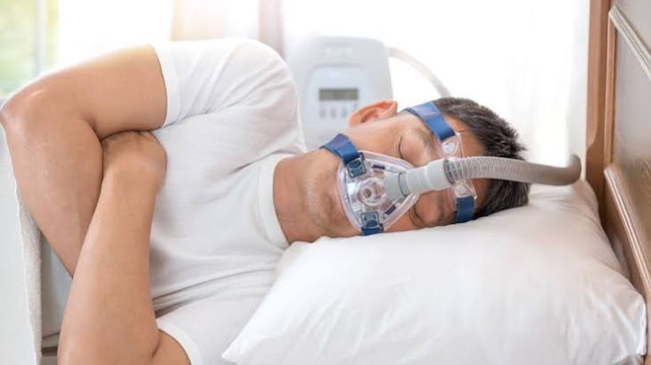
Continuous Positive Airway Pressure (CPAP): The gold standard treatment for obstructive sleep apnea is CPAP therapy, which delivers a gentle, continuous stream of pressurized air through a mask worn during sleep, keeping the upper airway propped open and preventing collapses.
-
Alternative Interventions: Depending on the underlying cause and severity, management may also include custom oral appliances designed to reposition the lower jaw, lifestyle modifications such as weight management and positional therapy, or surgical procedures to remove excess soft tissue or correct anatomical obstructions.
Conclusion
Sleep apnea is far more than a frustrating nighttime inconvenience marked by loud snoring; it is a serious, systemic medical disorder that inflicts profound stress on the central nervous system. By understanding how repetitive airway collapses trigger intermittent hypoxia, oxidative stress, neuroinflammation, and structural damage within vulnerable brain regions like the hippocampus and prefrontal cortex, individuals can better appreciate the neurological risks of untreated sleep. Through accurate diagnostic sleep studies and effective interventions like CPAP therapy, patients can restore healthy oxygenation, protect cognitive function, and safeguard long-term brain health.