How High Cholesterol Can Lead to Heart Disease
Cholesterol is a soft, waxy, fat-like substance that is absolutely essential for normal human biological functioning. Far from being a mere toxin, cholesterol is manufactured primarily by the liver and is required to build healthy cellular membranes, synthesize vital steroid hormones such as estrogen, testosterone, and cortisol, and produce bile acids necessary for the digestion of dietary fats. Because cholesterol is insoluble in blood, it cannot travel through the vascular system on its own; instead, it must bind to specialized proteins to form water-soluble complexes known as lipoproteins. However, when the delicate balance of these lipoproteins is disrupted—specifically when low-density lipoprotein cholesterol accumulates in excess—it sets off a pathological cascade that can culminate in severe cardiovascular disease. Understanding how high cholesterol systematically damages the arterial architecture provides vital insight into preventing heart attacks and strokes.
Lipoprotein Transport and the Distinction Between LDL and HDL
To comprehend how cholesterol drives heart disease, one must first examine the two primary classes of lipoproteins responsible for transporting cholesterol through the bloodstream: low-density lipoprotein and high-density lipoprotein.
-
Low-Density Lipoprotein (LDL): Often termed "bad" cholesterol, LDL is responsible for carrying cholesterol particles synthesized in the liver out to peripheral tissues and cells that require it for cellular repair and hormone production. When LDL levels are excessively high relative to the body's actual metabolic demands, these circulating particles linger in the bloodstream for extended periods, increasing their vulnerability to chemical modification and arterial deposition.
-
High-Density Lipoprotein (HDL): Conversely, HDL is known as "good" cholesterol because it performs a protective scavenging role. HDL particles travel through the vascular network, gathering excess, unneeded cholesterol from peripheral tissues and arterial walls and transporting it back to the liver for safe processing, degradation, and excretion from the body.
Endothelial Injury and the Initiation of Atherosclerosis
The pathogenesis of heart disease driven by high cholesterol begins silently within the innermost lining of the blood vessels, a delicate single layer of cells known as the endothelium.
-
The Protective Endothelium: A healthy endothelium regulates vascular tone, prevents unnecessary blood clotting, and acts as a selective barrier preventing circulating blood components from infiltrating the deep muscular walls of the arteries.
-
Vascular Insult and Permeability: Chronic exposure to high concentrations of circulating LDL cholesterol—compounded by cardiovascular risk factors like hypertension, cigarette smoking, and diabetes—causes chemical injury and oxidative stress to endothelial cells. This injury compromises the tight junctions between endothelial cells, making the vascular wall abnormally permeable and allowing small, dense LDL particles to slip past the surface barrier and lodge directly into the sub-endothelial space of the arterial wall.
Oxidation, Foam Cell Formation, and Fatty Streaks
Once trapped beneath the endothelial lining, the accumulated LDL particles undergo harmful chemical modifications that trigger an aggressive immune response.
-
Oxidative Modification: Free radicals and reactive oxygen species present within the arterial wall chemically alter the trapped LDL molecules, converting them into oxidized LDL.
-
Macrophage Recruitment: The body's immune system recognizes this oxidized material as foreign and dangerous, dispatching white blood cells known as monocytes to the site. These monocytes burrow into the arterial wall, mature into specialized scavenger cells called macrophages, and aggressively engulf the oxidized LDL particles.
-
Lipid Engorgement: Overwhelmed by the sheer volume of ingested cholesterol, these macrophages become swollen with fat droplets, transforming permanently into lipid-laden "foam cells." The microscopic accumulation of these foam cells creates the earliest visible sign of vascular disease: flat, yellowish fatty streaks lining the inner surface of the arteries.
Plaque Maturation and Fibrous Cap Development
As the inflammatory process persists over months and years, the initial fatty streak evolves into a complex, structurally sophisticated lesion known as an atherosclerotic plaque.
-
Smooth Muscle Proliferation: Chemical signals released by trapped immune cells stimulate smooth muscle cells from the middle layer of the artery to migrate toward the inner lining, where they multiply and secrete a tough, fibrous matrix of collagen and elastin.
-
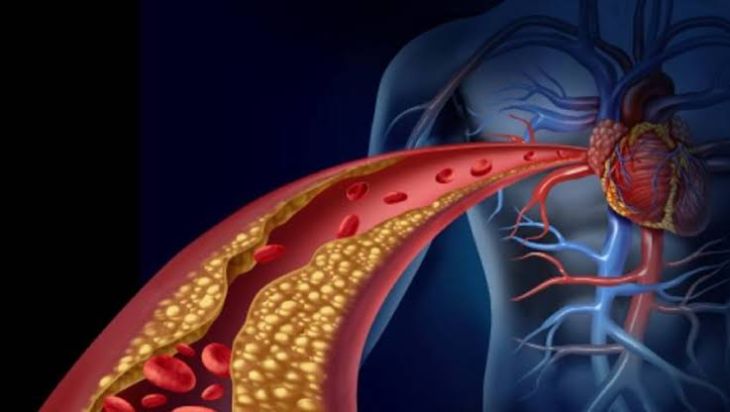
The Fibrous Cap: This fibrous tissue forms a protective cap over the underlying core of cellular debris, foam cells, and extracellular cholesterol crystals. Within the growing plaque, localized oxygen deprivation can cause cells in the center to die, forming a soft, necrotic lipid pool. The artery attempts to compensate for this narrowing by expanding outward initially, but eventually, the growing plaque encroaches directly upon the vessel lumen, progressively obstructing normal blood flow.
Ischemia, Angina, and Coronary Artery Disease
When atherosclerotic plaques restrict blood flow within the coronary arteries—the specialized blood vessels tasked with supplying oxygen-rich blood directly to the heart muscle itself—the clinical syndrome known as coronary artery disease (CAD) develops.
-
Myocardial Ischemia: Under resting conditions, a moderately narrowed coronary artery may still supply adequate blood. However, during periods of physical exertion or emotional stress—when the heart muscle demands an increased supply of oxygen—the narrowed vessel fails to deliver sufficient blood flow, resulting in a mismatch known as myocardial ischemia.
-
Angina Pectoris: This oxygen starvation manifests clinically as angina pectoris, a characteristic squeezing, pressure, or tightness in the chest that often radiates to the jaw, neck, or left arm, signaling that the heart muscle is struggling to function due to restricted perfusion.
Plaque Rupture, Thrombosis, and Acute Myocardial Infarction
While chronic vessel narrowing causes stable angina, the most catastrophic cardiovascular events—such as acute heart attacks—are frequently triggered by sudden, unpredictable changes in plaque stability.
-
Vulnerable Plaques: Not all plaques are hard and calcified; many dangerous plaques possess a large, soft lipid core covered by a dangerously thin, fragile fibrous cap weakened by ongoing inflammation.
-
Thrombus Formation: If this thin cap ruptures, the highly thrombogenic interior of the plaque is suddenly exposed to circulating blood. Platelets rush to the site, rapidly adhering to the rupture and triggering the formation of a massive blood clot (thrombus). This clot can completely occlude the coronary artery within minutes, cutting off all blood supply to a region of the heart muscle and causing permanent tissue death, or a myocardial infarction.
Conclusion
High cholesterol is a silent yet profoundly destructive driver of cardiovascular disease, operating through a well-documented pathophysiological sequence that transforms flexible arteries into rigid, narrowed conduits. By understanding how excess low-density lipoprotein infiltrates injured endothelium, undergoes oxidation, transforms macrophages into foam cells, and builds complex atherosclerotic plaques, the mechanics of heart disease become clear. Recognizing these cellular pathways highlights the critical importance of regular cholesterol screenings, lifestyle modifications, and targeted medical therapies designed to lower LDL levels, stabilize arterial plaques, and protect long-term heart health.