What Is Coronary Artery Disease and How Does It Develop?
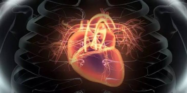
The human heart is an extraordinary muscular pump that beats approximately one hundred thousand times daily, circulating roughly two thousand gallons of blood to sustain every tissue in the body. Despite holding a constant volume of blood within its chambers, the heart muscle cannot extract oxygen or nutrients directly from this internal supply. Instead, the myocardium relies entirely on a dedicated, specialized network of blood vessels known as the coronary arteries, which branch directly off the base of the aorta to wrap around the heart like a crown. These vital conduits must deliver a continuous, high-volume supply of oxygen-rich blood, particularly during periods of physical exertion or emotional stress when the heart's metabolic demands escalate dramatically. However, when chronic pathological processes progressively narrow or obstruct these critical vessels, individuals develop coronary artery disease (CAD), the single leading cause of mortality and cardiovascular complications globally.
The Foundation of Vascular Health and Endothelial Integrity
To understand how coronary artery disease develops, one must first examine the micro-anatomical structure of a healthy coronary artery. The inner lining of these vessels consists of a delicate, single layer of specialized cells called the endothelium, which plays an active, highly regulated role in maintaining cardiovascular health. A healthy endothelium produces nitric oxide and other vasoactive molecules that regulate vascular tone, prevent unnecessary blood clotting, and act as a selective barrier preventing circulating blood components from infiltrating the deep muscular walls of the artery. Coronary artery disease begins when various systemic risk factors—such as chronic hypertension, dyslipidemia, cigarette smoking, and diabetes mellitus—inflict continuous chemical insult and physical stress upon this fragile endothelial lining, compromising its normal protective functions.
Endothelial Injury and Lipid Infiltration
The initial, silent phase of coronary artery disease development centers around structural injury and abnormal permeability within the coronary vessel wall.
-
Compromised Barrier Function: Chronic exposure to high blood pressure creates mechanical strain, while elevated circulating levels of low-density lipoprotein (LDL) cholesterol undergo chemical modification as they pass through the stressed endothelium.
-
Sub-Endothelial Deposition: Because the injured endothelial cells lose their tight intercellular junctions, small, dense LDL particles slip past the surface barrier and lodge directly into the sub-endothelial space of the arterial wall. Once trapped within this intimal layer, the LDL molecules are chemically altered by reactive oxygen species, becoming oxidized LDL. This oxidized material triggers a localized immune response, signaling the body to dispatch white blood cells called monocytes to the site of injury in an attempt to clear away the perceived threat.
Foam Cell Formation and Fatty Streak Development
As the inflammatory response escalates, the immune system's cellular response inadvertently drives the progression of the underlying vascular disease.
-
Macrophage Ingestion: The recruited monocytes burrow through the endothelium, mature into aggressive scavenger cells known as macrophages, and ingest the trapped, oxidized LDL particles in large quantities.
-
Foam Cell Accumulation: Overwhelmed by the sheer volume of ingested fat, these macrophages swell into lipid-laden "foam cells." The microscopic aggregation of these foam cells creates the earliest visible sign of coronary artery disease: a flat, yellowish fatty streak lining the inner surface of the coronary artery. While fatty streaks can appear even in children and young adults, they represent the foundational cellular nidus from which complex, obstructive atherosclerotic plaques eventually evolve over decades.
Plaque Progression and Fibrous Cap Formation
What begins as a microscopic accumulation of foam cells gradually transforms into a sophisticated, structurally complex lesion known as an atherosclerotic plaque.
-
Smooth Muscle Migration: Chemical signaling molecules released by trapped immune cells stimulate smooth muscle cells from the middle layer of the artery to migrate toward the inner lining, where they multiply and secrete a tough, fibrous matrix of collagen and elastin.
-
Necrotic Core Creation: This fibrous tissue forms a protective cap over a growing core of cellular debris, dead foam cells, and extracellular cholesterol crystals. As this plaque expands, it thickens the arterial wall and begins to encroach upon the vessel lumen, slowly restricting the cross-sectional area through which blood can flow to the heart muscle.
Myocardial Ischemia and the Onset of Clinical Symptoms
While early plaque growth is often accommodated by outward expansion of the coronary artery, eventually the lesion becomes large enough to impede blood flow significantly, precipitating clinical symptoms.
-
Supply-Demand Mismatch: Under resting conditions, a moderately narrowed coronary artery may still supply adequate blood. However, during physical exertion or stress, the heart demands more oxygen, but the stenotic vessel fails to deliver sufficient perfusion, resulting in myocardial ischemia.
-
Angina Pectoris: This oxygen starvation manifests as angina pectoris—a characteristic chest pressure, tightness, or squeezing that often radiates to the left arm, jaw, or neck—signaling that the heart muscle is struggling to function under restricted blood flow.
Plaque Rupture, Thrombosis, and Acute Myocardial Infarction
The most dangerous phase of coronary artery disease involves sudden, unpredictable changes in plaque stability rather than slow, steady narrowing.
-
Vulnerable Plaques: Many dangerous plaques possess a large, soft lipid core covered by a thin, fragile fibrous cap weakened by ongoing inflammation.
-
Acute Thrombosis: If this thin cap ruptures, the highly thrombogenic interior of the plaque is suddenly exposed to circulating blood. Platelets rush to the site, rapidly adhering to the rupture and triggering the formation of a massive blood clot that can completely occlude the coronary artery within minutes, cutting off all blood supply to a region of the heart muscle and causing a myocardial infarction.
Conclusion
Coronary artery disease is a progressive, chronic cardiovascular condition driven by atherosclerosis, wherein lipid accumulation, endothelial injury, and chronic inflammation transform flexible coronary arteries into narrowed, obstructed conduits. By examining the step-by-step pathological sequence—from initial LDL infiltration and foam cell formation to fibrous cap maturation and acute plaque rupture—the complex mechanisms of heart disease become clear. Recognizing these cellular pathways highlights the critical importance of early cardiovascular screenings, rigorous lifestyle modifications, and targeted medical therapies designed to lower cholesterol, control blood pressure, and protect long-term heart health.