How Pancreatic Disorders Can Affect Digestion and Blood Sugar
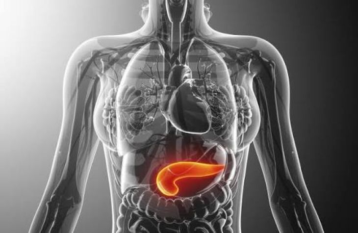
The human pancreas is a remarkable, elongated gland situated deep within the upper abdomen, tucked behind the stomach and adjacent to the small intestine. Despite its modest size, the pancreas serves as a metabolic powerhouse, uniquely designed to perform two completely distinct yet equally vital physiological roles essential for human survival. It functions as both an exocrine gland, manufacturing and secreting powerful digestive enzymes into the gastrointestinal tract, and an endocrine gland, producing critical hormones that regulate systemic blood sugar levels. When acute inflammation, chronic structural damage, autoimmune destruction, or neoplastic growths compromise this delicate glandular architecture, individuals develop complex pancreatic disorders that simultaneously sabotage nutrient digestion and destabilize glucose homeostasis. Understanding how the pancreas governs these dual systems illuminates the profound metabolic consequences of pancreatic disease.
The Exocrine Pancreas and the Mechanics of Nutrient Digestion
To understand how pancreatic disorders disrupt digestion, one must first examine the normal exocrine function of the gland. Roughly ninety-five percent of the pancreas is dedicated to exocrine activity, consisting of specialized clusters of cells called acinar cells that synthesize and secrete inactive digestive enzymes into a network of small ducts. These ducts converge into the main pancreatic duct, which delivers enzyme-rich pancreatic juice directly into the duodenum—the first section of the small intestine.
-
Enzymatic Breakdown: This secretion contains vital enzymes tailored to break down every major macronutrient: amylase digests complex carbohydrates into simple sugars, lipases dismantle dietary fats into absorbable fatty acids and monoglycerides, and proteases like trypsin and chymotrypsin cleave proteins into small peptides and amino acids.
-
Bicarbonate Neutralization: Additionally, ductal cells secrete large volumes of bicarbonate, a powerful alkaline buffer that neutralizes corrosive gastric acid entering the small intestine from the stomach, creating the optimal pH environment required for these digestive enzymes to function effectively.
The Endocrine Pancreas and Blood Sugar Homeostasis
In stark contrast to the exocrine network, the endocrine pancreas consists of scattered clusters of specialized cells known as the islets of Langerhans, which make up the remaining five percent of the glandular mass. These microscopic endocrine islands act as the body's primary master thermostat for glucose regulation, releasing hormones directly into the bloodstream.
-
Insulin Secretion: When blood glucose levels rise following a meal, beta cells within the islets secrete insulin, a hormone that unlocks cellular doors throughout the body, allowing circulating glucose to enter muscle, fat, and liver cells to be utilized for immediate energy or stored for later use.
-
Glucagon Counter-Regulation: Conversely, when blood sugar drops between meals or during fasting, alpha cells release glucagon, signaling the liver to break down stored glycogen into glucose to maintain steady, life-sustaining energy supplies for the brain and vital organs.
Acute Pancreatitis and Sudden Metabolic Disruptions
Acute pancreatitis is a sudden, severe inflammation of the pancreas most commonly triggered by gallstones blocking the pancreatic duct or excessive alcohol consumption.
-
Premature Enzyme Activation: Normally, digestive enzymes remain inactive until they reach the small intestine; however, in acute pancreatitis, cellular injury causes these powerful enzymes to activate prematurely while still trapped inside the pancreas.
-
Autodigestion: This triggers a destructive process known as autodigestion, where the pancreas begins to digest its own tissue, causing excruciating upper abdominal pain, systemic inflammation, and profound physiological shock. During acute attacks, the sudden destruction of both acinar and islet cells can cause temporary exocrine failure—leading to malabsorption and severe steatorrhea—alongside acute hyperglycemia or dangerously fluctuating blood sugar levels as insulin-producing beta cells are damaged by the inflammatory storm.
Chronic Pancreatitis and Progressive Glandular Destruction
When acute inflammation recurs repeatedly or persists over many years, it leads to chronic pancreatitis, a progressive fibrotic disease characterized by the permanent scarring and destruction of functional pancreatic tissue.
-
Fibrosis and Calcification: As chronic inflammation destroys acinar and islet cells, normal glandular tissue is systematically replaced by dense, fibrous scar tissue, often accompanied by calcification within the pancreatic ducts.
-
Exocrine Insufficiency: Over time, this loss of functional mass results in pancreatic exocrine insufficiency (PEI), a debilitating condition where the pancreas can no longer produce enough enzymes to digest food. Without sufficient lipase and proteases, fats and proteins pass unabsorbed through the digestive tract, resulting in chronic diarrhea, severe abdominal cramping, foul-smelling oily stools, and profound malnutrition despite a normal or ravenous appetite.
Pancreatogenic Diabetes and Endocrine Failure
As chronic pancreatitis or advanced pancreatic cancer destroys the structural architecture of the pancreas, the endocrine islets of Langerhans are inevitably caught in the destructive path, leading to a distinct form of secondary diabetes known as type 3c diabetes or pancreatogenic diabetes.
-
Loss of Beta and Alpha Cells: Unlike type 1 diabetes, which is an autoimmune condition, or type 2 diabetes, which involves insulin resistance, type 3c diabetes stems from the direct physical loss of both insulin-producing beta cells and glucagon-producing alpha cells.
-
Brittle Glucose Control: This dual loss creates a uniquely challenging clinical picture characterized by "brittle" diabetes, where patients experience wild, unpredictable swings between dangerously low blood sugar and severe hyperglycemia. Because alpha cells are also destroyed, the body lacks the counter-regulatory glucagon response needed to rescue the patient from hypoglycemia, making blood sugar management exceptionally difficult and requiring meticulous insulin therapy and specialized nutritional support.
Diagnostic Evaluations and Clinical Investigation
Diagnosing complex pancreatic disorders requires a comprehensive clinical evaluation combining detailed patient histories, laboratory testing, and advanced imaging modalities.
-
Biomarker Analysis: Gastroenterologists and endocrinologists utilize blood tests to measure elevated levels of pancreatic enzymes—such as serum amylase and lipase—alongside assessments of long-term blood sugar control via glycated hemoglobin (HbA1c) and fasting glucose levels. To evaluate exocrine function directly, physicians analyze stool samples for fecal elastase, a protein that indicates whether the pancreas is producing adequate digestive enzymes.
-
Advanced Imaging: Furthermore, high-resolution diagnostic imaging—including abdominal computed tomography (CT) scans, magnetic resonance cholangiopancreatography (MRCP), and endoscopic ultrasound (EUS)—allows specialists to visualize structural scarring, ductal obstructions, calcifications, and masses with extraordinary precision, guiding targeted medical or surgical interventions.
Conclusion
Pancreatic disorders encompass destructive conditions such as acute and chronic pancreatitis that compromise the dual exocrine and endocrine functions of the gland, simultaneously sabotaging nutrient digestion and destabilizing blood sugar regulation. By examining how acinar cell destruction leads to fat malabsorption and exocrine insufficiency, and how islet cell scarring precipitates brittle pancreatogenic diabetes, the profound metabolic impact of pancreatic disease becomes clear. Through thorough diagnostic testing, precise enzyme replacement therapy, meticulous blood sugar management, and advanced medical interventions, healthcare providers can mitigate nutritional deficiencies, stabilize glucose homeostasis, and significantly improve the quality of life for affected patients.