Understanding Common Pancreatic Diseases and Their Warning Signs
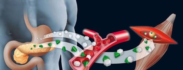
The human pancreas is a deeply situated, elongated retroperitoneal organ nestled snugly within the upper abdomen behind the stomach, intimately linked with the duodenum and major vascular structures. Despite its modest size, the pancreas serves as a vital cornerstone of human metabolism through its unique dual role as both an exocrine and an endocrine gland. The exocrine pancreas comprises the vast majority of the organ's mass, consisting of specialized acinar cells and ductal networks that synthesize, store, and secrete powerful digestive enzymes—including trypsinogen, lipase, and amylase—into the small intestine to break down proteins, fats, and carbohydrates. Simultaneously, scattered throughout the exocrine tissue are microscopic clusters of cells known as the islets of Langerhans, which constitute the endocrine pancreas. These specialized cells secrete critical hormones directly into the bloodstream, most notably insulin and glucagon, which orchestrate systemic glucose homeostasis. When inflammatory cascades, autodigestive injury, or malignant transformations disrupt this delicate architecture, individuals face complex pancreatic diseases that profoundly impact digestion and metabolic control.
Acute Pancreatitis: Autodigestion and Systemic Inflammatory Response
One of the most common and acutely painful conditions affecting the organ is acute pancreatitis, an inflammatory disorder characterized by the sudden activation of digestive enzymes within the pancreatic parenchyma itself. Under normal circumstances, digestive enzymes remain safely inactive until they reach the lumen of the small intestine. However, when pathological triggers induce premature intracellular activation of trypsin, these powerful enzymes begin digesting the pancreas's own cellular proteins and membranes, a destructive process known as pancreatic autodigestion. This localized injury triggers an intense inflammatory response, recruiting neutrophils and macrophages, causing microvascular thrombosis, and leading to interstitial edema, localized cell death, and occasionally hemorrhagic necrosis. The primary clinical drivers of acute pancreatitis are gallstones—which can temporarily lodge in the ampulla of Vater and obstruct pancreatic duct outflow—and chronic, excessive alcohol consumption. Patients present with a sudden onset of severe, constant epigastric pain that frequently radiates straight through to the back, accompanied by persistent nausea, vomiting, and abdominal tenderness.
Chronic Pancreatitis: Progressive Fibrosis and Exocrine-Endocrine Failure
While acute pancreatitis represents a sudden, potentially reversible inflammatory event, repeated bouts of acute inflammation or continuous low-grade injury can permanently alter the structural integrity of the organ, leading to chronic pancreatitis. Chronic pancreatitis is characterized by progressive, irreversible destruction of the pancreatic parenchyma, accompanied by extensive fibrosis, hardening of the tissue, and stricturing or calcification of the pancreatic ducts. Over time, the acinar cells responsible for producing digestive enzymes are progressively replaced by scar tissue, resulting in exocrine pancreatic insufficiency where the body can no longer produce adequate enzymes to digest food properly. This manifests clinically as steatorrhea—foul-smelling, fatty, difficult-to-flush stools—alongside profound weight loss and malnutrition. Furthermore, as the chronic fibrotic scarring encroaches upon the islets of Langerhans, endocrine function is similarly compromised, eventually leading to secondary diabetes mellitus characterized by brittle blood sugar fluctuations.
Pancreatic Cancer: Pathological Transformation and Early Detection Challenges
Among all malignancies affecting the digestive system, pancreatic ductal adenocarcinoma stands out as one of the most aggressive and lethal forms of cancer, arising most frequently from the epithelial cells lining the small pancreatic ducts. The pathological development of pancreatic cancer is driven by a series of cumulative genetic mutations that transform normal ductal epithelium into invasive, rapidly proliferating neoplastic tissue. This aggressive cancer is notorious for its insidious onset; in its early stages, tumors grow deep within the retroperitoneal space without impinging upon vital neighboring structures, allowing the disease to advance silently before triggering noticeable symptoms. Consequently, the vast majority of patients are diagnosed at an advanced, unresectable stage, underscoring the critical need to identify subtle early warning signs and understand the specific anatomical locations of tumors within the head, body, or tail of the pancreas.
Critical Warning Signs and Clinical Symptom Presentation
Recognizing the early warning signs of pancreatic disease is exceptionally challenging because symptoms frequently overlap with common gastrointestinal complaints, yet certain clinical red flags demand urgent investigation.
-
Abdominal Pain and Back Radiation: Persistent, dull epigastric pain that bores straight through to the mid-back and is occasionally relieved by leaning forward is a hallmark feature of both chronic pancreatitis and pancreatic malignancies involving the body or tail of the organ.
-
Painless Jaundice: Tumors located specifically in the head of the pancreas often compress the common bile duct early in their progression, obstructing the flow of bile into the intestine and causing painless jaundice—manifesting as yellowing of the skin and sclera, accompanied by dark, tea-colored urine and pale, clay-colored stools.
-
Constitutional Symptoms: Unexplained, profound weight loss coupled with sudden-onset muscle wasting, new-onset atypical diabetes mellitus in an older adult without traditional risk factors, and debilitating fatigue serve as critical systemic warning signs that warrant immediate cross-sectional abdominal imaging.
Diagnostic Investigations and Advanced Imaging Modalities
Diagnosing complex pancreatic diseases requires sophisticated diagnostic modalities capable of visualizing an organ hidden deep behind the stomach and transverse colon.
-
Cross-Sectional Imaging: Advanced multidetector computed tomography (CT) scans utilizing specialized pancreatic protocols serve as the primary diagnostic workhorse, providing high-resolution cross-sectional views to evaluate parenchymal inflammation, necrosis, ductal dilation, and tumor margins. Magnetic resonance cholangiopancreatography offers a non-invasive method to map the biliary and pancreatic ductal trees with exceptional clarity.
-
Endoscopic Ultrasound: Furthermore, endoscopic ultrasound—where a specialized ultrasound probe is advanced through the stomach directly adjacent to the pancreas—allows gastroenterologists to visualize tiny lesions, assess vascular invasion, and perform fine-needle aspirations to obtain tissue samples for definitive histological diagnosis.
Therapeutic Interventions and Comprehensive Management
Managing pancreatic diseases requires a coordinated, multidisciplinary approach tailored entirely to the specific pathology and stage of the condition. Acute pancreatitis is managed primarily through aggressive supportive care in a hospital setting, focusing on rigorous intravenous fluid resuscitation to maintain end-organ perfusion, aggressive pain management, and nutritional support, alongside endoscopic retrograde cholangiopancreatography to remove obstructing gallstones when indicated. Chronic pancreatitis requires long-term lifestyle modifications—including strict abstinence from alcohol and smoking cessation—coupled with pancreatic enzyme replacement therapy to manage malabsorption and targeted insulin regimens for diabetes. For localized pancreatic malignancies, complex surgical interventions such as the Whipple procedure offer the only potential for long-term cure, though they are frequently combined with systemic chemotherapy and radiation therapy to optimize outcomes and extend survival.
Conclusion
Pancreatic diseases encompass a spectrum of challenging conditions—ranging from acute and chronic pancreatitis to aggressive ductal adenocarcinoma—that fundamentally impair both exocrine digestion and endocrine metabolic control. By examining how autodigestion, progressive fibrosis, and malignant cellular transformations disrupt pancreatic architecture, the vital importance of this retroperitoneal organ becomes clear. Through meticulous clinical evaluation of warning signs like painless jaundice and radiating back pain, advanced cross-sectional imaging, endoscopic diagnostics, and targeted medical and surgical interventions, healthcare providers can successfully manage inflammation, alleviate chronic symptoms, and improve survival outcomes for affected individuals.