How Could Microfluidic Chips Help Researchers Study Diseases Using Tiny Samples?
Studying diseases often requires blood samples, tissue samples, or other biological materials. Traditional laboratory tests may need specialized equipment, larger sample volumes, and several steps to produce results.
Microfluidic chips are changing how researchers approach some of these tasks. These small devices contain tiny channels that control the movement of liquids, allowing scientists to study cells, proteins, and other biological materials using very small samples.
What Are Microfluidic Chips?
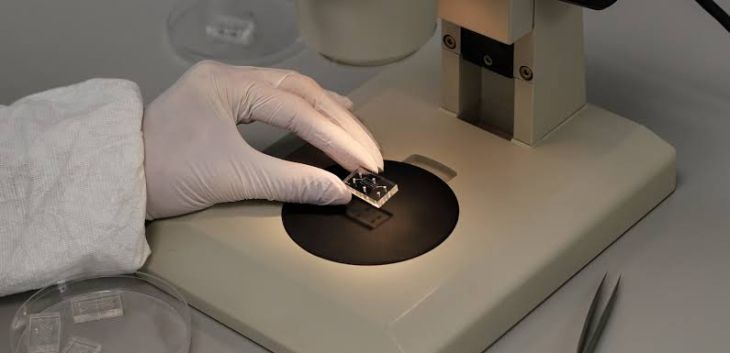
Microfluidic chips are small devices designed to handle extremely small amounts of liquid through narrow channels. These channels can be thinner than a strand of hair and allow researchers to control how fluids move, mix, and react.
The devices may be made from materials such as glass, plastic, or specialized polymers. Some contain sensors, tiny valves, or chambers that help scientists perform specific experiments.
Microfluidic chips are sometimes called lab-on-a-chip devices because they can combine several laboratory processes within one small system.
For example, a chip may be designed to separate cells from a blood sample, detect certain molecules, or observe how cells respond to a medicine.
How Do Microfluidic Chips Work?
Microfluidic chips use small channels and chambers to move and control liquids. Researchers introduce a sample into the device and guide it through different areas where specific processes take place.
The general process includes several steps:
Sample collection: Researchers collect a small amount of blood, saliva, or another biological sample.
Sample preparation: The sample may be processed to separate cells, proteins, or other substances of interest.
Movement through tiny channels: The liquid travels through the chip's internal pathways.
Testing and measurement: Sensors or imaging systems detect biological signals or changes.
Data analysis: Researchers study the results to understand what the sample reveals about a disease or biological process.
The exact process depends on the chip's design and the research question.
How Can Microfluidic Chips Help Researchers Study Diseases?
Microfluidic technology can help scientists investigate diseases at a detailed level while using less biological material.
Studying Individual Cells
Many diseases involve changes in how cells grow, communicate, or respond to their surroundings. Microfluidic chips can isolate or control individual cells, allowing researchers to examine their behavior under specific conditions.
This can be valuable when studying immune cells, cancer cells, and other cell populations that behave differently during disease.
Analyzing Blood Samples
Blood contains many types of cells, proteins, and other substances that may provide information about a person's health.
Some microfluidic devices are designed to identify particular cells or molecules in small blood samples. Researchers are studying how these systems could help detect disease-related changes without requiring large sample volumes.
However, a research device is not automatically a clinically approved diagnostic test. Each application needs appropriate testing to establish its accuracy.
Investigating Infections
Microfluidic chips can be designed to detect biological materials associated with certain bacteria or viruses. Researchers are exploring these systems for faster testing and more efficient analysis of infectious diseases.
Some platforms may combine sample preparation and detection in a single device, reducing the number of laboratory steps required.
Their real-world performance depends on the type of infection, sample quality, and detection method.
Could Microfluidic Chips Improve Cancer Research?
Cancer research is one of the promising applications of microfluidic technology.
Cancer cells can differ from one another, even within the same tumor. Some may grow quickly, spread more easily, or respond differently to treatment.
Microfluidic chips can help researchers study these differences by creating controlled environments for cancer cells. Scientists can expose cells to different medicines and observe how they respond.
Researchers are also exploring ways to capture rare cells circulating in blood, including certain tumor cells. These methods may help investigate how cancer spreads and how tumors change over time.
Although these approaches are promising, they do not replace established cancer screening or diagnostic methods unless a particular test has been properly validated for clinical use.
What Is an Organ-on-a-Chip System?
An organ-on-a-chip is a specialized type of microfluidic device designed to imitate certain features of human organs.
For example, researchers can grow human cells inside tiny channels and expose them to controlled fluid flow or other conditions. Some devices aim to recreate important features of lung, liver, heart, or intestinal tissue.
These systems can help scientists study how diseases affect tissues and how medicines behave in environments that resemble parts of the human body.
Organ-on-a-chip models may also reduce the need for some early experiments involving animals. However, they cannot yet reproduce every function and interaction found in a complete human organ.
What Are the Main Benefits of Microfluidic Chips?
Microfluidic chips offer several potential advantages in medical research:
Smaller sample requirements: Many designs work with tiny amounts of biological material.
Controlled experiments: Researchers can carefully manage fluid movement and testing conditions.
Faster analysis: Combining laboratory steps may reduce the time needed for some experiments.
Reduced reagent use: Small volumes can lower the amount of expensive chemicals or testing materials required.
Detailed cell studies: Certain chips allow researchers to observe how individual cells respond to different conditions.
Portable testing possibilities: Some designs may eventually enable testing outside traditional laboratories.
These benefits vary by device and application, and not every chip offers all of them.
What Challenges Do Researchers Face?
Despite their potential, microfluidic chips have limitations. Designing and manufacturing reliable devices can be difficult, particularly when they need to handle complex biological samples.
Tiny channels may become blocked, and results can be affected by bubbles, sample quality, or differences in operating conditions. Some devices also require specialized sensors, imaging equipment, or trained staff.
Another challenge is translating laboratory results into dependable medical tests. Researchers must show that a device produces accurate, consistent results across different samples and settings before it can be used for routine clinical decisions.
What Is the Future of Microfluidic Technology?
Scientists are combining microfluidic chips with artificial intelligence, advanced sensors, and cell-based models to improve disease research.
These combinations may help researchers analyze small samples, identify biological patterns, and test potential medicines more efficiently. In the future, certain technologies could contribute to faster diagnostic tests and more personalized approaches to treatment.
However, many applications remain under development. Their benefits will depend on careful validation, practical costs, and evidence that they work reliably in real medical settings.
FAQs
Can microfluidic chips detect diseases?
Some microfluidic devices can detect specific biological markers associated with disease. Whether a device can be used for diagnosis depends on its design, accuracy, and clinical validation.
Do microfluidic chips require blood samples?
Not always. Depending on the application, researchers may use blood, saliva, other bodily fluids, or cells grown in a laboratory.
How do microfluidic chips help cancer research?
They allow researchers to study cancer cells under controlled conditions, investigate medicine responses, and explore ways to detect rare tumor-related cells in blood.
What is the difference between a microfluidic chip and an organ-on-a-chip?
A microfluidic chip is a broad category of devices that control tiny fluid volumes. An organ-on-a-chip is a specialized system that uses microfluidics and living cells to imitate selected features of an organ.
Are microfluidic chips used in hospitals?
Some microfluidic technologies are used in diagnostic and laboratory applications, while many newer systems remain experimental. Their availability depends on the specific device and its approved use.
Conclusion
Microfluidic chips are opening new ways to study diseases using very small biological samples. They can help researchers examine individual cells, analyze blood, investigate infections, and test potential treatments in controlled environments. Although challenges remain in manufacturing, reliability, and clinical validation, continued advances may make these devices increasingly valuable in medical research. By combining small-scale fluid control with modern sensors and data analysis, microfluidic technology could contribute to more efficient disease studies and future diagnostic innovations.