Understanding Brain Aneurysms and Their Warning Signs
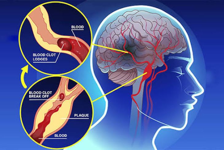
A brain aneurysm—clinically referred to as a cerebral or intracranial aneurysm—is a localized, abnormal structural weakness in the wall of an artery located within the cranial vault. Much like a thin, over-stretched spot on an inflating balloon, this vulnerable zone can gradually expand over time due to the relentless, high-pressure force of systemic blood circulation. While numerous small aneurysms remain clinically silent and produce no adverse health complications throughout an individual's lifetime, others present significant, life-altering medical concerns if they enlarge, leak, or undergo sudden structural failure. Because these vascular conditions frequently develop without external physical indicators until a critical threshold is reached, studying their underlying physiological mechanics and identifying potential warning signals is essential for timely clinical intervention. A severe rupture constitutes an urgent medical crisis involving rapid, pressurized bleeding into the surrounding cerebrospinal fluid pathways, demanding immediate neurosurgical care to mitigate the risk of permanent neurological impairment or fatality.
The Vascular Mechanics: How Arterial Weakness Develops
Intracranial bulges typically form at branching junctions along the major arteries supplying oxygenated blood to the brain. These specific bifurcation points—most notably within the Circle of Willis—experience continuous, focused mechanical stress from the pulsing flow of blood. Medical researchers have identified several primary, overlapping factors contributing to the gradual breakdown of arterial wall integrity. Chronic hemodynamic turbulence at sharp vascular curves gradually wears down the internal endothelial and muscular layers of the vessel, while some individuals are born with intrinsically thinner muscular structures in their arterial walls, increasing their susceptibility to long-term stretching.
Furthermore, sustained, unmanaged high blood pressure forces blood against these vessel walls with excessive velocity, accelerating microscopic wear and tear. Beyond these physical stressors, specific genetic traits affecting collagen and connective tissue elasticity can further compromise the overall resilience of the circulatory network, making the development of these bulges a multifaceted interaction between genetics and systemic health. Conditions such as autosomal dominant polycystic kidney disease, Ehlers-Danlos syndrome, and familial history heavily influence an individual's lifetime baseline risk profile, requiring heightened clinical vigilance among susceptible populations.
Identifying Indicators of an Unruptured Vascular Bulge
The vast majority of small, intact vascular bulges do not generate any noticeable bodily sensations, as they often exist without compressing adjacent structures. However, if a bulge expands sufficiently in size or location, it may apply localized physical pressure against nearby cranial nerves or delicate neural tissue, producing distinct "sentinel" red flags. Individuals might experience persistent, deep, or sharp orbital discomfort positioned directly behind one eye, or notice the sudden onset of a dilated pupil that fails to react normally to changes in light.
Additional clinical signs include the sudden drooping of the upper eyelid, known as ptosis, or the development of double vision resulting from localized nerve compression caused by pressure on the third cranial nerve. Furthermore, patients may report unusual tingling or numbness restricted to one side of the face, or frequent, intense head pain that differs notably in intensity, frequency, and character from their historical headache patterns. When these specific neurological markers present themselves, consulting a neurologist for advanced magnetic resonance angiography or computed tomography imaging is the standard, necessary clinical procedure to rule out an enlarging vascular anomaly before a catastrophic failure occurs.
Critical Warning Indicators of an Acute Rupture
A sudden rupture of an intracranial artery wall is a life-threatening medical emergency. When the structural boundary of the aneurysm fails, pressurized blood rapidly escapes into the subarachnoid space, causing an instantaneous and dangerous surge in internal cranial pressure. Immediate emergency medical assistance must be contacted if symptoms appear, beginning with the primary hallmark of an explosive, instantaneous thunderclap headache. Often described by survivors as the most severe head pain they have ever experienced, this discomfort reaches its peak intensity within seconds of onset and is completely distinct from standard tension headaches or chronic migraines.
This event is frequently accompanied by projectile, intense nausea and severe vomiting triggered by the rapidly escalating pressure inside the skull. Other classic emergency indicators include severe nuchal rigidity, where neck stiffness becomes so pronounced that it is physically impossible to touch the chin to the chest, and ocular photophobia, manifesting as an agonizing, painful intolerance to normal ambient light levels. Additional warning signs include sudden, profound confusion, disorientation, temporary loss of consciousness, and the abrupt onset of convulsive seizure activity in an adult who has no prior history of epilepsy or neurological disorders.
Diagnostic Evaluation Methods
Clinical specialists utilize a variety of sophisticated non-invasive and minimally invasive imaging technologies to map cerebral vasculature with high precision. In emergency department settings, non-contrast computed tomography scans are frequently utilized as the first line of defense to quickly identify the presence of fresh blood accumulation within the cranial cavity. Following a positive scan, physicians often order a computed tomography angiography, which utilizes an intravenous contrast medium to generate detailed, three-dimensional representations of the brain’s entire arterial tree, allowing medical teams to pinpoint the exact location and geometry of the vascular defect.
Magnetic resonance angiography provides a valuable alternative by employing magnetic fields and radio frequencies to visualize blood vessels without exposure to ionizing radiation. For definitive pre-surgical planning, clinicians may also utilize digital subtraction angiography, a specialized catheter-based procedure that provides high-resolution, real-time visualization of blood flow dynamics, allowing surgeons to see exactly how blood enters and exits the bulge before mapping out a definitive operational strategy.
Modern Therapeutic Interventions
Medical strategies for managing these conditions are highly customized based on the exact dimensions, shape, and anatomical positioning of the bulge, as well as the patient’s overall cardiovascular health status and age. In traditional surgical clipping, a neurosurgeon performs a craniotomy to access the base of the bulge, applying a small, permanent titanium clip to physically seal it off from normal blood circulation, effectively excluding the sac from arterial pressure permanently.
Endovascular coiling offers a contemporary, minimally invasive alternative where a micro-catheter is guided through the vascular system from the femoral artery to place soft platinum coils into the aneurysm cavity. These coils induce a natural, localized clotting response that effectively isolates the weak zone from arterial pressure without requiring an open skull procedure. Alternatively, flow diversion devices utilize specialized, fine-mesh stents placed across the neck of larger or more complex bulges to reroute blood flow smoothly through the parent artery, which encourages the body to naturally remodel and heal the vessel wall over time.
Long-Term Vascular Health Maintenance
While certain innate genetic characteristics cannot be modified, adopting rigorous heart-healthy habits helps reduce overall cardiovascular strain and protects the integrity of the vascular system. Maintaining blood pressure metrics strictly within medically recommended ranges through consistent daily monitoring and prescribed antihypertensive therapies is vital for those at risk. Eliminating tobacco use is perhaps the most impactful lifestyle change, as nicotine and other chemicals in smoke act as primary catalysts for arterial wall degradation, endothelial inflammation, and plaque accumulation.
Emphasizing a diet rich in whole foods, leafy green vegetables, healthy fats, and adequate hydration while actively moderating daily sodium intake supports long-term vascular health. Additionally, engaging in consistent, moderate aerobic physical activity helps maintain endothelial function and overall circulatory resilience, further lowering the cumulative stress placed on the brain’s delicate arterial network. Patients should also maintain open, ongoing communication with their primary care providers regarding any new neurological shifts.
Conclusion
Intracranial vascular bulges require careful medical awareness, yet proactive health management can significantly influence long-term clinical outcomes. Recognizing subtle, early-stage neurological warnings and treating sudden, severe thunderclap headaches with absolute emergency urgency remains the most vital step in ensuring patient safety. Through the combination of advanced diagnostic imaging and modern, minimally invasive therapeutic techniques, medical professionals can effectively address these complex vascular conditions, prevent catastrophic ruptures, and support sustained, lifelong neurological well-being. By prioritizing cardiovascular health and maintaining regular check-ups with primary care physicians, individuals can take definitive control over their neurovascular future and safeguard their long-term health.